


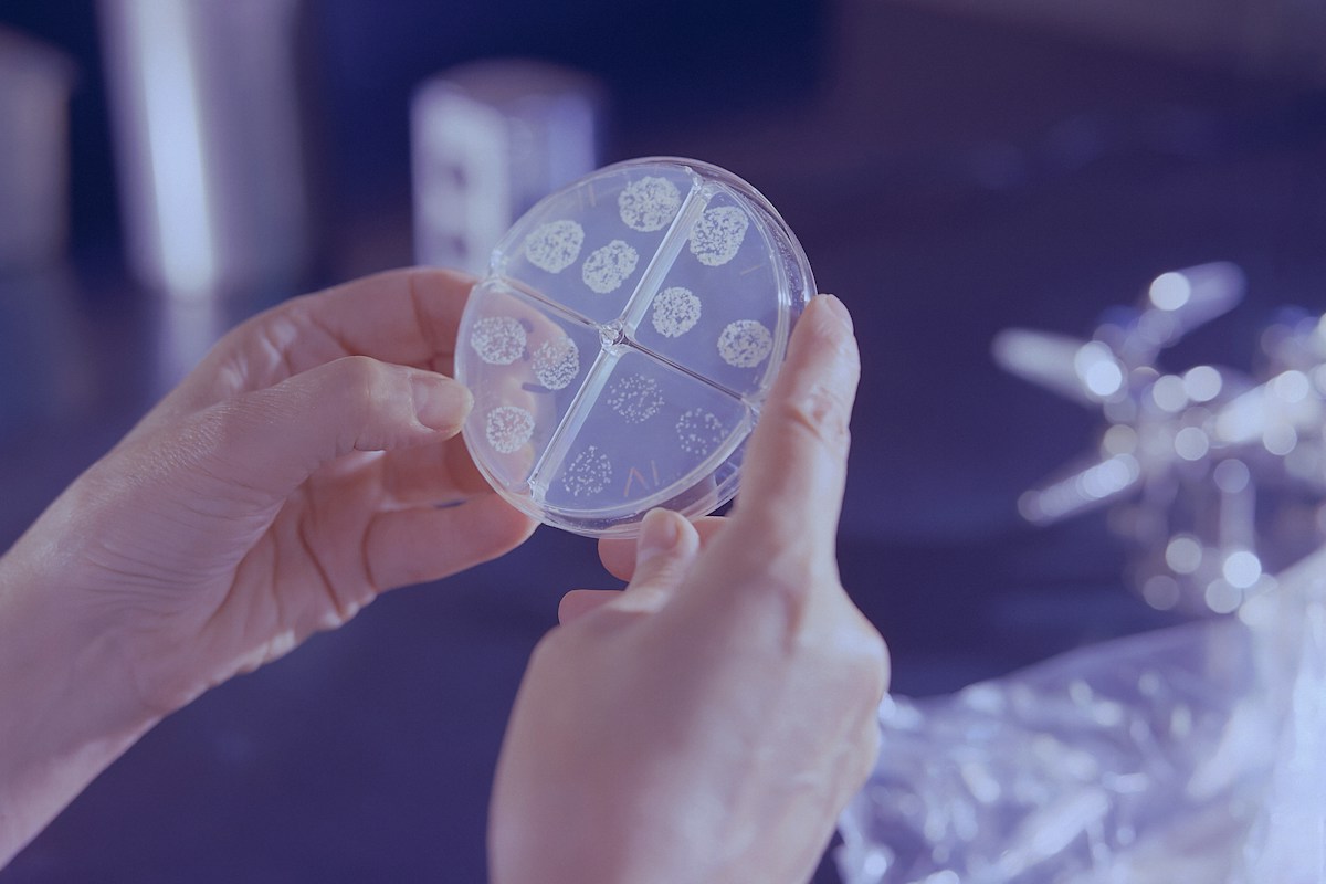
The human gastrointestinal tract harbors an immensely complex, dense ecological consortium of over 100 trillion microbial organisms comprising bacteria, archaea, fungi, protozoa, and bacteriophages, collectively encoding over 3 million unique genes. Historically regarded as passive commensals restricted to nutrient digestion and fecal bulk generation, contemporary biomedical science recognizes the gut microbiome as an endocrine, metabolic, and neuroimmunological powerhouse. Through continuous bi-directional communication networks collectively designated the gut-brain-immune axis, the gut microbiota orchestrates Central Nervous System (CNS) development, neural circuit refinement, blood-brain barrier integrity, and adult neurogenesis.
The primary biochemical currency mediating this cross-talk consists of microbial metabolites, most prominently short-chain fatty acids (SCFAs) – acetate, propionate, and butyrate – produced via the anaerobic fermentation of non-digestible dietary prebiotic fibers by specialized anaerobic commensals. Operating both locally in the intestinal mucosa and systemically via the portal and systemic circulations, SCFAs cross the blood-brain barrier via monocarboxylate transporters, binding G-protein coupled receptors (GPR41/FFAR3, GPR43/FFAR2, GPR109A) and inhibiting class I and class II histone deacetylases (HDACs). This epigenetic and signaling axis directly modulates the maturation, morphological ramification, and synaptic pruning functions of microglia – the resident parenchymal macrophage sentinels of the mammalian central nervous system.
This comprehensive clinical research treatise investigates the mechanistic architecture of the gut-brain-immune axis. We dissect the biophysical pathways connecting the enteric nervous system to the brainstem via the vagus nerve, examine how microbial dysbiosis triggers systemic lipopolysaccharide endotoxemia and neuroinflammation, evaluate microglial phenotypic polarization in neurodegenerative diseases such as Parkinson’s and Alzheimer’s, and examine translational therapeutic frontiers including targeted psychobiotics, personalized prebiotic synbiotics, and fecal microbiota transplantation.
The Architecture of the Gut-Brain-Immune Axis: Tripartite Communication
The gut-brain-immune axis represents a multidirectional, organ-spanning communication network integrating the gastrointestinal tract, the enteric nervous system (ENS), the central nervous system (CNS), the autonomic nervous system (ANS), the hypothalamic-pituitary-adrenal (HPA) axis, and systemic innate and adaptive immune networks.
This tripartite network operates through three distinct yet synchronized modalities: neural pathways mediated by the afferent and efferent branches of the vagus nerve and spinal sympathetic nerves; endocrine pathways mediated by gut hormone peptide secretion and adrenal corticosteroids; and immune-metabolic pathways mediated by bacterial metabolites, cell wall antigens, and cytokine cascades.
Within the gut wall itself lies the enteric nervous system, frequently termed the ‘second brain’. Comprising over 500 million intrinsic neurons organized into the myenteric (Auerbach’s) and submucosal (Meissner’s) plexuses, the ENS functions semi-autonomously to control gastrointestinal motility, mucosal secretion, microvascular perfusion, and local immune defenses.
Crucially, the enteric nervous system is separated from the dense luminal microbial biomass by a single monolayer of specialized intestinal epithelial cells (IECs) and an overlying mucus barrier. This micro-anatomical arrangement places immune and neural sensors in immediate physical contact with microbial metabolites.
When mucosal homeostasis is preserved, the microbiota trains the immune system to maintain oral tolerance while providing neurotrophic signals that foster central neuroplasticity and emotional resilience. Conversely, disruptions in microbial ecology ignite a vicious cycle of mucosal permeability, systemic endotoxemia, and neuroinflammation.
Microbial Metabolomics: Anaerobic Fermentation and Short-Chain Fatty Acid Synthesis
The primary metabolic output of the colonic microbiota is the fermentation of complex dietary carbohydrates, resistant starches, and soluble plant fibers that escape enzymatic digestion in the human stomach and small intestine.
Colonic anaerobic commensals – predominantly belonging to the bacterial phyla Bacillota (formerly Firmicutes) and Bacteroidota (formerly Bacteroidetes) – utilize specialized carbohydrate-active enzymes (CAZymes) to hydrolyze these complex glycans into simple hexoses and pentoses, subsequently fermenting them into short-chain fatty acids (SCFAs).
The three most abundant SCFAs produced in the human colon are acetate (C2), propionate (C3), and butyrate (C4), which typically occur in a molar ratio of approximately 60:20:20 in healthy individuals, reaching total luminal concentrations exceeding 100 to 140 mM in the proximal colon.
Acetate is synthesized primarily by members of the Bacteroidetes phylum and Bifidobacterium species via the Wood-Ljungdahl pathway or the acetyl-CoA pathway; propionate is synthesized predominantly by Bacteroides and Veillonella via the succinate or acrylate pathways; and butyrate is produced by specialized Clostridial clusters IV and XIVa (including Faecalibacterium prausnitzii, Eubacterium rectale, and Roseburia spp.) via the butyryl-CoA:acetate CoA-transferase pathway.
Once produced, SCFAs are rapidly absorbed across the colonic apical membrane via passive diffusion or electrogenic sodium-coupled monocarboxylate transporters (SMCT1 / SLC5A8) and proton-coupled monocarboxylate transporters (MCT1 / SLC16A1).
Butyrate: Epigenetic Master Regulator and Histone Deacetylase Inhibition
Among all microbial metabolites, butyrate occupies a unique physiological status as the primary bioenergetic substrate for colonocytes and a master epigenetic modifier across mammalian tissues.
In healthy colonic epithelial cells, butyrate is transported into mitochondria, where it undergoes beta-oxidation to acetyl-CoA, feeding the citric acid cycle and generating over 70 percent of the total ATP consumed by the intestinal epithelium. This rapid metabolic consumption maintains physiological hypoxia at the mucosal surface, preserving an anaerobic lumen essential for obligate anaerobe survival.
Beyond its role as a cellular fuel, butyrate functions as an endogenous, potent non-selective inhibitor of class I and class II histone deacetylases (HDACs). By inhibiting HDAC activity at sub-millimolar concentrations, butyrate prevents the removal of acetyl groups from the lysine tails of histone proteins H3 and H4.
This increases histone acetylation, opening chromatin into a transcriptionally permissive euchromatin conformation. In colonic mucosal regulatory T cells (Tregs), butyrate-mediated HDAC inhibition upregulates the transcription factor FOXP3, driving the differentiation and proliferation of anti-inflammatory, interleukin-10-secreting Tregs.
Systemically, butyrate enters the portal and peripheral circulations, crossing the blood-brain barrier to inhibit HDACs within central microglia and neurons, where it upregulates brain-derived neurotrophic factor (BDNF), enhances synaptic plasticity, and represses pro-inflammatory cytokine transcription.
Receptor-Mediated Signaling: FFAR2, FFAR3, and GPR109A Transduction Cascades
In addition to intracellular epigenetic modulation, short-chain fatty acids function as high-affinity ligands for specific cell-surface G-protein coupled receptors: Free Fatty Acid Receptor 2 (FFAR2 / GPR43), Free Fatty Acid Receptor 3 (FFAR3 / GPR41), and Hydroxycarboxylic Acid Receptor 2 (HCA2 / GPR109A).
FFAR2 exhibits highest affinity for acetate and propionate, coupling to both Gi/o and Gq/11 signaling pathways. Expressed broadly on intestinal epithelial cells, neutrophils, monocytes, and dendritic cells, FFAR2 activation stimulates intracellular calcium mobilization and activates mitogen-activated protein kinase (MAPK) cascades, orchestrating immune cell recruitment, mucosal healing, and regulatory immune maturation.
FFAR3 is selectively activated by propionate and butyrate, coupling exclusively to Gi/o proteins to inhibit adenylate cyclase and decrease intracellular cyclic AMP (cAMP) levels. FFAR3 is densely expressed on enteric neurons, autonomic sympathetic ganglia, and peripheral sensory neurons, where it directly modulates gut peristalsis, sympathetic nervous outflow, and metabolic rate.
GPR109A is highly selective for butyrate (and the endogenous hepatic ketone body beta-hydroxybutyrate). Expressed on colonic epithelial cells, macrophages, and microglia, GPR109A activation exerts potent anti-inflammatory effects by suppressing nuclear factor kappa-light-chain-enhancer of activated B cells (NF-kB) translocation and inhibiting inflammasome assembly.
Through these cell-surface receptors, luminal microbial metabolites transmit immediate biochemical information to the vascular, nervous, and immune systems without requiring massive systemic concentrations.
Vagus Nerve: The Rapid Neuroanatomical Conduit
The vagus nerve (tenth cranial nerve, CN X) represents the most direct, rapid bidirectional neuroanatomical highway connecting the gastrointestinal tract to the brainstem. Approximately 80 to 90 percent of the vagal nerve fibers traversing the diaphragm are afferent (sensory) fibers transmitting visceral information from the gut directly to the nucleus tractus solitarius (NTS) in the medulla oblongata.
Vagal afferent nerve terminals terminate within the mucosal lamina propria and the muscularis externa of the stomach and intestine, forming intricate intraganglionic laminar endings. While vagal sensory terminals do not cross the epithelial barrier into the bacterial lumen, they are positioned in immediate proximity to enteroendocrine cells (EECs) and mucosal dendritic cells.
Enteroendocrine cells – such as neuropod-bearing enterochromaffin cells and L-cells – directly sense microbial metabolites and bacterial flagellin via apical receptors. In response to SCFA stimulation, EECs release neurotransmitters and peptide hormones, including serotonin (5-hydroxytryptamine / 5-HT), cholecystokinin (CCK), glucagon-like peptide-1 (GLP-1), and peptide YY (PYY).
These signaling molecules bind specific receptors on adjacent vagal afferent nerve terminals, depolarizing the nerve membrane and transmitting high-frequency sensory action potentials directly to the brainstem within milliseconds.
From the nucleus tractus solitarius, visceral sensory signals project to the parabrachial nucleus, locus coeruleus, hypothalamus, amygdala, and insular cortex, profoundly modulating mood, anxiety, autonomic tone, appetite, and central neuroinflammatory states.
The Cholinergic Anti-Inflammatory Pathway: Efferent Neuro-Immune Suppression
Communication along the vagus nerve is not purely sensory; efferent vagal motor fibers originating in the dorsal motor nucleus of the vagus (DMV) execute the ‘cholinergic anti-inflammatory pathway’, a powerful mechanism discovered by Kevin Tracey that suppresses systemic inflammation.
When afferent vagal signals notify the brainstem of peripheral inflammatory cascades or endotoxemia, the DMV dispatches efferent action potentials down the vagus nerve. Because the vagus nerve does not directly innervate the spleen, these efferent fibers terminate in the celiac-superior mesenteric ganglion plexus.
From this ganglion, postganglionic sympathetic fibers of the splenic nerve travel into the red and white pulp of the spleen, releasing norepinephrine in close proximity to a specialized subset of memory T lymphocytes (CD4+ CD44high CD62Llow T cells) that express choline acetyltransferase (ChAT).
Upon beta-2 adrenergic receptor stimulation by norepinephrine, these ChAT-positive T cells synthesize and release the neurotransmitter acetylcholine (ACh) into the splenic parenchyma.
Acetylcholine binds alpha-7 nicotinic acetylcholine receptors (alpha-7 nAChR) expressed on splenic macrophages. Alpha-7 nAChR signaling suppresses phosphorylation of IkB-alpha, preventing NF-kB nuclear translocation and blocking the synthesis and secretion of tumor necrosis factor-alpha (TNF-alpha), interleukin-1beta (IL-1beta), and interleukin-6 (IL-6), effectively dampening peripheral cytokine storms.
Blood-Brain Barrier Integrity and Microbial Regulation
The blood-brain barrier (BBB) is a specialized, tightly sealed multicellular neurovascular interface comprising brain capillary endothelial cells, pericytes, and the end-feet of astrocytes. The BBB strictly restricts the paracellular diffusion of hydrophilic solutes, macromolecules, toxins, and peripheral immune cells into the cerebral parenchyma, preserving central biochemical homeostasis.
Endothelial sealing is executed by continuous inter-endothelial junctional complexes composed of tight junction proteins – including claudin-5, occludin, and zonula occludens-1 (ZO-1) – anchored to the actin cytoskeleton.
Landmark investigations conducted in germ-free (GF) mice – animals born and raised in sterile isolators devoid of any microorganisms – revealed that the microbiome is indispensable for the structural establishment and lifelong maintenance of blood-brain barrier integrity.
Germ-free mice exhibit marked, continuous hyperpermeability of the blood-brain barrier from embryonic development through adulthood, characterized by disorganized, severely downregulated expression of claudin-5 and occludin throughout brain microvessels.
Remarkably, when germ-free mice are monocolonized with butyrate-producing bacterial strains or administered physiological concentrations of sodium butyrate, claudin-5 and occludin expression is fully restored within days, re-establishing tight junction integrity and sealing the blood-brain barrier against systemic circulating macromolecules.
Microglia: The Central Nervous System Parenchymal Macrophages
Microglia constitute approximately 10 to 15 percent of all cells in the human brain, functioning as the primary resident innate immune sentinels and housekeeping phagocytes of the central nervous system parenchyma.
Unlike peripheral tissue macrophages that derive from bone marrow hematopoiesis, microglia possess a unique developmental origin: they derive from early erythro-myeloid progenitors (EMPs) in the embryonic yolk sac, migrating into the developing neural tube prior to the formation of the blood-brain barrier, where they self-renew throughout life.
In the healthy adult brain, ‘resting’ or homeostatic microglia are not dormant; their cell bodies remain anchored while their extensive, highly ramified, motile cytoplasmic processes continuously survey the brain parenchyma, palpating surrounding synapses, astrocytes, and blood vessels every few hours.
Homeostatic microglia express a unique transcriptomic signature governed by the transcription factor Sall1, characterized by surface expression of purinergic receptors (P2RY12), fractalkine receptor (CX3CR1), and transmembrane protein 119 (TMEM119).
These homeostatic sentinels clear metabolic debris, release neurotrophic factors (BDNF, IGF-1), and actively monitor synaptic firing patterns to support cognitive function and emotional equilibrium.
Microglial Pruning and Synaptic Refinement: Developmental and Adult Homeostasis
During critical developmental windows, the embryonic and neonatal brain overproduces synaptic connections. To sculpt functional, precise neural circuits, excess, weak, or inactive synapses must be selectively identified and eliminated – a process termed synaptic pruning.
Microglia execute synaptic pruning primarily through the classical complement cascade. Inactive or redundant presynaptic terminals and dendritic spines are tagged with the complement initiator proteins C1q and C3.
Microglia express complement receptor 3 (CR3, an integrin complex of CD11b and CD18). Upon binding C3b opsonized onto synaptic terminals, microglial processes engulf the synapse via phagocytosis, transporting it into lysosomal compartments for enzymatic destruction.
Astonishingly, Erny and colleagues demonstrated in a landmark 2015 study that the gut microbiota is required for normal microglial development and synaptic pruning capacity. In germ-free mice, microglia exhibit profound morphological defects: their cell bodies are enlarged, their ramified processes are stunted, and they express immature, blunted transcriptomic profiles with impaired innate immune responsiveness.
Recolonization of germ-free mice with complex, diverse microbiota or oral administration of a physiological cocktail of SCFAs (acetate, propionate, butyrate) fully rescues microglial maturation, restores normal ramified morphology, and normalizes synaptic pruning activity, proving that the gut microbiome continuously programs central macrophage maturation.
Intestinal Dysbiosis, Epithelial Barrier Decay, and Leaky Gut Syndrome
When the delicate equilibrium of the colonic microbial ecosystem is disrupted – secondary to ultra-processed diets, chronic psychological stress, repeated broad-spectrum antibiotic courses, or environmental toxins – the state of dysbiosis emerges.
Dysbiosis is characterized by three cardinal features: the loss of microbial alpha-diversity, the depletion of protective, obligate anaerobic taxa (such as Faecalibacterium prausnitzii, Bifidobacteria, and Akkermansia muciniphila), and the pathological bloom of pro-inflammatory, facultative anaerobic pathobionts, primarily within the Enterobacteriaceae family.
Depleted of dietary fermentable fibers and butyrate-producing anaerobes, the colonic mucus layer thins dramatically as starving bacteria consume host mucin glycoproteins for survival. Furthermore, colonic epithelial tight junction proteins (claudin-1, occludin, ZO-1) dissociate secondary to cellular energetic starvation and localized oxidative stress.
This pathological breakdown in epithelial barrier function – clinically referred to as increased intestinal permeability or ‘leaky gut’ – permits the unhindered paracellular translocation of luminal contents into the lamina propria and mesenteric venules.
The most clinically consequential consequence of this barrier collapse is metabolic endotoxemia: the continuous trans-mucosal translocation of Gram-negative bacterial lipopolysaccharide (LPS) directly into the systemic circulation.
Metabolic Endotoxemia: Toll-Like Receptor 4 Activation and Cytokine Cascades
Lipopolysaccharide (LPS), also termed endotoxin, is a major glycolipid component of the outer membrane of Gram-negative bacteria. LPS molecules consist of a hydrophobic Lipid A domain, an oligosaccharide core, and a repeating O-antigen polysaccharide chain.
Under healthy baseline conditions, minimal LPS traces penetrate the mucosal barrier, being swiftly bound by circulating lipopolysaccharide-binding protein (LBP) and cleared by hepatic Kupffer cells via scavenger receptors. In the setting of dysbiosis and mucosal hyperpermeability, portal clearance is overwhelmed, and circulating plasma endotoxin concentrations rise 2- to 3-fold above baseline, establishing chronic metabolic endotoxemia.
Circulating LPS binds Toll-Like Receptor 4 (TLR4) complexes on monocytes, dendritic cells, and endothelial cells in conjunction with coreceptors CD14 and MD-2. TLR4 dimerization engages the adaptor proteins MyD88 and TRIF, initiating downstream phosphorylation of the IkappaB kinase (IKK) complex.
Active IKK phosphorylates IkappaB, targeting it for proteasomal degradation and freeing the transcription factor NF-kB to translocate into the cell nucleus. Nuclear NF-kB drives intense transcription of pro-inflammatory cytokines: TNF-alpha, IL-1beta, IL-6, and monocyte chemoattractant protein-1 (MCP-1 / CCL2).
These circulating cytokines travel via the bloodstream to cerebral microvessels, where they bind receptors on brain endothelial cells, initiating downstream inflammatory signaling cascades across the blood-brain barrier.
Neuroinflammation: Microglial Polarization from Homeostatic to Reactive States
Within the central nervous system, persistent systemic inflammatory signals and elevated circulating LPS trigger the phenotypic transformation of homeostatic microglia into reactive, neurodestructive states.
Circulating pro-inflammatory cytokines stimulate brain capillary endothelial cells to release prostaglandins (PGE2) and nitric oxide into the brain parenchyma. In regions where the blood-brain barrier is naturally fenestrated – the circumventricular organs such as the area postrema and median eminence – LPS can directly enter the brain tissue.
Upon exposure to LPS, TNF-alpha, or damaged neuronal membranes, microglia undergo a profound morphological and functional shift: they retract their delicate, ramified processes, enlarge their somas, and adopt an amoeboid, migratory morphology reminiscent of activated macrophages.
Reactive microglia downregulate homeostatic markers (P2RY12, TMEM119) and upregulate pro-inflammatory markers including CD68, MHC class II, inducible nitric oxide synthase (iNOS), and cyclooxygenase-2 (COX-2). They assemble the NLRP3 inflammasome, producing high concentrations of reactive oxygen species (ROS), peroxynitrite, IL-1beta, and TNF-alpha.
In this reactive state, microglia lose their supportive synaptic maintenance functions and begin indiscriminately engulfing viable neuronal synapses, inducing profound synaptic loss, inhibiting hippocampal adult neurogenesis, and triggering neurodegenerative cascades.
The Microbiome in Parkinson’s Disease: Braak’s Hypothesis and Alpha-Synuclein
Parkinson’s disease (PD) represents the clearest clinical and neuropathological illustration of the gut-to-brain pathological progression in humans.
Over two decades ago, German neuropathologist Heiko Braak formulated the groundbreaking hypothesis that Parkinson’s disease does not originate within the brain, but rather initiates in the gut, triggered by an unknown pathogen or environmental toxin that induces protein misfolding in enteric nerves.
The pathognomonic histological hallmark of Parkinson’s disease is the intracellular accumulation of insoluble Lewy bodies composed predominantly of misfolded, phosphorylated alpha-synuclein protein aggregates. In Parkinson’s patients, gastrointestinal dysfunction – particularly severe constipation – precedes motor symptoms (tremor, rigidity, bradykinesia) by 10 to 20 years.
Biopsies of the gastrointestinal tract in early-stage PD patients reveal phosphorylated alpha-synuclein inclusions within the submucosal and myenteric plexuses. Experimental animal models have proven that misfolded alpha-synuclein fibrils injected into the colonic wall propagate retrogradely in a prion-like manner along the vagus nerve to the dorsal motor nucleus of the vagus in the brainstem, ultimately ascending to the substantia nigra.
Critically, patients who have undergone complete truncal vagotomy (surgical severing of the vagus nerve) for peptic ulcer disease decades earlier exhibit a dramatic 40 to 50 percent reduction in their subsequent lifetime risk of developing Parkinson’s disease, confirming the vagus nerve as the structural conduit of pathology.
The Microbiome in Alzheimer’s Disease: Amyloid Cross-Seeding and Neurodegeneration
Emerging clinical and translational research has firmly linked gut microbial dysbiosis to the pathogenesis and progression of Alzheimer’s disease (AD), the leading cause of dementia worldwide.
Metagenomic sequencing of fecal samples from Alzheimer’s patients reveals severe microbial dysbiosis characterized by significant reductions in butyrate-producing taxa (such as Butyrivibrio, Eubacterium, and Roseburia) and marked enrichment of pro-inflammatory Gram-negative taxa (Bacteroides and Escherichia).
Many intestinal bacterial species – including Escherichia coli, Salmonella enterica, Pseudomonas aeruginosa, and Klebsiella pneumoniae – naturally produce functional extracellular bacterial amyloids (such as curli fibers, CsgA, and Fap proteins) to form protective structural biofilms.
These bacterial amyloids share biophysical quaternary structural motifs with human amyloid-beta (Abeta1-42) and hyperphosphorylated tau. Exposure of the intestinal mucosa and systemic immune system to bacterial amyloids can trigger ‘cross-seeding’: bacterial amyloid fibrils act as nucleating templates that accelerate the misfolding and aggregation of endogenous human Abeta in the brain through molecular mimicry.
Concurrently, chronic LPS endotoxemia downregulates low-density lipoprotein receptor-related protein 1 (LRP1) at the blood-brain barrier – the primary efflux transporter responsible for pumping Abeta out of the brain into the circulation – leading to massive cerebral Abeta retention and senile plaque formation.
Psychiatric Consequences: Major Depression, Anxiety, and the HPA Axis
Beyond classical neurodegenerative disorders, the gut-brain-immune axis plays a profound role in the neurobiology of major depressive disorder (MDD) and generalized anxiety disorder.
Depression and anxiety are increasingly understood not merely as monoaminergic neurochemical imbalances, but as systemic conditions characterized by chronic, low-grade neuroinflammation and hypothalamic-pituitary-adrenal (HPA) axis hyper-reactivity.
In the brain, chronic microglial activation and elevated pro-inflammatory cytokines (TNF-alpha, IL-6) induce the enzyme indoleamine 2,3-dioxygenase (IDO-1) in microglia and astrocytes. IDO-1 shunts dietary L-tryptophan away from the synthesis of serotonin (5-HT) and melatonin, diverting it instead into the kynurenine pathway.
Downstream metabolism of kynurenine by activated microglia generates quinolinic acid – a potent, excitotoxic agonist of the N-methyl-D-aspartate (NMDA) receptor that causes dendritic spine loss, glutamate excitotoxicity, and hippocampal neuronal atrophy.
Simultaneously, microbial dysbiosis impairs glucocorticoid receptor feedback sensitivity in the hypothalamus, locking the HPA axis into a continuous state of hyper-cortisolemia that further erodes hippocampal neurogenesis and impairs prefrontal emotional regulation.
The Enteric Nervous System: Intrinsic Myenteric and Submucosal Circuitry Dynamics
The enteric nervous system (ENS) represents an extraordinarily complex, self-contained neural network embedded directly within the tissue wall of the gastrointestinal tract, stretching from the proximal esophagus to the internal anal sphincter.
Containing roughly 500 million neurons – a population greater than the entire spinal cord – the ENS is organized into two major concentric ganglionated plexuses: the outer myenteric plexus (Auerbach’s plexus), positioned between the longitudinal and circular smooth muscle layers to control motor peristalsis, and the inner submucosal plexus (Meissner’s plexus), situated adjacent to the lamina propria to regulate local mucosal blood flow, epithelial fluid secretion, and mucosal electrolyte transport.
The ENS utilizes more than thirty distinct neurotransmitters and neuropeptides identical to those found in the brain, including acetylcholine, substance P, vasoactive intestinal peptide (VIP), nitric oxide, dopamine, and serotonin. Primary afferent neurons (intrinsic primary afferent neurons, or IPANs) have sensory terminals projecting into the lamina propria, where they monitor mechanical distension, chemical osmolarity, and luminal microbial metabolites.
When luminal SCFAs like acetate and propionate bind FFAR3 on enteric sensory neurons, they directly modulate action potential frequency, fine-tuning migrating motor complexes (MMCs) and propagating peristaltic contractions. Conversely, microbial pathobionts that secrete neurotoxic peptides can inhibit IPAN firing, inducing profound enteric neuropathy, intestinal pseudo-obstruction, or dysmotility syndromes.
This rich intrinsic circuitry enables the ENS to orchestrate complex digestive reflexes completely independently of CNS input, while continuously transmitting processed visceral sensory intelligence upward to the central nervous system via vagal and spinal pathways.
Tryptophan Catabolism and Indole Signaling: Microbial AhR Ligands and Mucosal Homeostasis
Dietary L-tryptophan is an essential aromatic amino acid that serves as a common substrate for three competing metabolic pathways: the serotonin synthesis pathway in enterochromaffin cells, the host kynurenine pathway in immune and hepatic cells, and direct microbial catabolism within the colonic lumen.
Commensal bacteria – notably including Lactobacillus species, Clostridium sporogenes, and Peptostreptococcus russellii – express tryptophanase and related metabolic enzymes that directly convert luminal tryptophan into diverse bioactive indole derivatives, including indole-3-propionic acid (IPA), indole-3-aldehyde (IAld), indole-3-acetic acid (IAA), and indoxyl sulfate.
Indole-3-propionic acid is an exceptionally potent, brain-penetrant natural antioxidant and neuroprotective agent. IPA scavenges hydroxyl radicals without generating pro-oxidant intermediates, suppresses beta-amyloid fibrillogenesis, and prevents microglial oxidative stress.
Crucially, microbial indoles function as high-affinity endogenous ligands for the Aryl Hydrocarbon Receptor (AhR) – an evolutionarily ancient ligand-activated transcription factor expressed abundantly on intestinal epithelial cells, innate lymphoid cells type 3 (ILC3s), and central astrocytes.
Activation of mucosal AhR by microbial IAld stimulates ILC3s to secrete interleukin-22 (IL-22). IL-22 binds epithelial receptors, inducing the expression of antimicrobial peptides (such as REG3gamma and calprotectin) and tight junction proteins, directly reinforcing the mucosal barrier and preventing pathogen translocation into systemic circulation.
Translational Therapeutics: Psychobiotics, Synbiotics, and Precision Dietary Interventions
Recognizing the decisive role of the microbiome in neurological and psychiatric disorders has opened a revolutionary therapeutic paradigm: microbial therapeutics designed to modify central brain function.
The term ‘psychobiotics’ was coined by John Cryan and Ted Dinan to define live biotherapeutic microorganisms that, when ingested in adequate amounts, confer mental health benefits by modulating gut-brain signaling. Specific strains – including Bifidobacterium longum 1714, Lactobacillus rhamnosus (JB-1), and Bifidobacterium infantis – have demonstrated profound neuro-modulatory efficacy in clinical trials.
Mechanistically, psychobiotics downregulate HPA axis stress responses, reduce circulating cortisol levels, increase plasma tryptophan bioavailability, stimulate enteric production of gamma-aminobutyric acid (GABA), and upregulate BDNF expression in the hippocampus and prefrontal cortex.
Precision synbiotics combine targeted probiotic strains with specific prebiotic prebiotic substrates (such as galacto-oligosaccharides [GOS], fructo-oligosaccharides [FOS], and partially hydrolyzed guar gum) to selectively nourish butyrate-producing taxa, driving sustained SCFA production.
Furthermore, dietary patterns rich in diverse fermentable fibers and polyphenols (such as the Mediterranean diet) consistently demonstrate protective effects against neurodegenerative and mood disorders by enhancing microbial alpha-diversity and preserving mucosal barrier integrity.
Fecal Microbiota Transplantation and Future Clinical Horizons
The most comprehensive approach to microbiome remodeling is Fecal Microbiota Transplantation (FMT): the transfer of a complete, screened microbial community from a healthy donor into the gastrointestinal tract of a patient.
While FMT is currently FDA-approved primarily for recurrent Clostridioides difficile infection, randomized clinical trials are exploring FMT as a groundbreaking investigational therapy for neurological conditions, including Parkinson’s disease, autism spectrum disorder, and treatment-resistant major depression.
Early clinical trial data demonstrate that FMT in Parkinson’s patients can substantially relieve both motor deficits and severe gastrointestinal symptoms, accompanied by marked reductions in serum neurofilament light chain (NfL) and pro-inflammatory cytokines.
Looking forward, the clinical frontier is moving from whole-stool transplantation toward defined, synthetic microbial consortia: precisely cultured, multi-strain bacterial cocktails designed to deliver specific metabolic outputs, such as high-volume butyrate synthesis and anti-inflammatory polysaccharide A expression.
Coupled with high-throughput metabolomics and single-cell microglial transcriptomics, precision microbiome medicine is poised to transform clinical neurology and psychiatry from organ-bound specialties into holistic, ecosystem-based disciplines.
| Signaling Pathway / Axis | Primary Molecular Mediators | Cellular Receptors & Targets | Central Nervous System Effects | Clinical & Pathology Correlates |
|---|---|---|---|---|
| Short-Chain Fatty Acids (SCFAs) | Acetate, Propionate, Butyrate | GPR41, GPR43, GPR109A, Class I/II HDACs | Microglial maturation, synaptic pruning, BDNF upregulation | Preservation of blood-brain barrier, anti-neuroinflammatory tone |
| Metabolic Endotoxemia | Gram-negative Lipopolysaccharide (LPS) | TLR4 / CD14 / MD-2 complex, MyD88, NF-kB | Microglial transformation to amoeboid reactive state, ROS/IL-1b release | Neurodegeneration, cognitive impairment, depressive anhedonia |
| Vagus Nerve Afferent Highway | Serotonin (5-HT), CCK, GLP-1, PYY from EECs | 5-HT3R, CCK1R, GLP-1R on vagal sensory nodose terminals | Direct visceral sensory input to NTS, limbic & cortical projection | Autonomic control, satiety, rapid mood & anxiety modulation |
| Kynurenine Excitotoxic Shunt | Quinolinic acid, Kynurenine, 3-hydroxykynurenine | NMDA receptors (NR2A/NR2B), Indoleamine 2,3-dioxygenase (IDO-1) | Excitotoxic dendritic spine loss, hippocampal atrophy, 5-HT deficit | Treatment-resistant depression, bipolar disorder, suicidality |
| Bacterial Amyloid Cross-Seeding | Curli fibers, CsgA, Fap bacterial biofilm proteins | Cross-seeding beta-sheet templates, TLR2/TLR1 | Accelerated aggregation of human Abeta1-42 & alpha-synuclein | Parkinson’s disease Lewy body ascendance, Alzheimer’s senile plaques |
The comparative matrix above synthesizes the major biological pathways operating within the gut-brain-immune axis. By examining the distinct biochemical mediators, receptor systems, and central physiological consequences across each pathway, clinicians can dissect how peripheral intestinal events drive complex central neurodegenerative and psychiatric manifestations.
Integrating these molecular pathways into clinical practice empowers healthcare practitioners to develop multi-modal therapeutic strategies that target both luminal microbial ecosystems and central neuroinflammatory cascades concurrently, bridging gastroenterology, neurology, and psychiatry.
Frequently Asked Questions Regarding the Gut-Brain-Immune Axis and Microglia
How do short-chain fatty acids cross the blood-brain barrier?
Short-chain fatty acids (SCFAs) – primarily acetate, propionate, and butyrate – cross the blood-brain barrier through specialized monocarboxylate transporters (MCTs), predominantly MCT1 (SLC16A1) and MCT4, which are densely expressed on brain capillary endothelial cells. In addition, uncharged, protonated forms of lipophilic SCFAs can traverse endothelial plasma membranes via passive non-ionic diffusion at physiological cerebral blood flow rates.
What is the primary difference between homeostatic and reactive microglia?
Homeostatic microglia exhibit a ramified morphology with small cell bodies and long, continuously motile branching processes that survey the neural parenchyma, expressing specific markers such as P2RY12, TMEM119, and CX3CR1. In contrast, reactive microglia undergo profound morphological transformation: they retract their processes, adopt an amoeboid, macrophage-like shape, downregulate homeostatic genes, and upregulate CD68, MHC-II, iNOS, and pro-inflammatory cytokines, actively producing reactive oxygen species and neurotoxic mediators.
Can the gut microbiota influence blood-brain barrier permeability?
Yes. Seminal research in germ-free mice has demonstrated that the absence of gut microbiota results in profound blood-brain barrier hyperpermeability due to downregulated expression of endothelial tight junction proteins, including claudin-5 and occludin. Colonization of germ-free animals with butyrate-producing bacteria or systemic administration of physiological sodium butyrate completely restores tight junction protein expression and re-establishes barrier integrity.
How does intestinal dysbiosis contribute to metabolic endotoxemia?
Intestinal dysbiosis involves the depletion of protective obligate anaerobes and the overgrowth of Gram-negative pathobionts. In the absence of fermentable dietary fiber, bacteria degrade the host colonic mucus layer. Concurrently, loss of butyrate impairs colonocyte tight junctions, increasing epithelial permeability. This allows lipopolysaccharide (LPS) from Gram-negative bacterial outer membranes to leak into the portal and systemic circulation, triggering widespread low-grade inflammation.
What is the cholinergic anti-inflammatory pathway?
The cholinergic anti-inflammatory pathway is a brain-to-immune reflex arc mediated by the vagus nerve. When the brainstem detects systemic inflammation, efferent vagal signals travel to the celiac ganglion and stimulate the splenic nerve. Noradrenaline released in the spleen stimulates specialized choline acetyltransferase-positive T cells to produce acetylcholine. Acetylcholine then binds alpha-7 nicotinic acetylcholine receptors on macrophages, shutting down NF-kB activation and halting the release of TNF-alpha, IL-1beta, and IL-6.
What are psychobiotics and which bacterial strains have been clinically validated?
Psychobiotics are specific probiotic bacterial strains that, when ingested in adequate therapeutic dosages, exert positive neurochemical and behavioral effects on mental health. Clinically validated strains include Bifidobacterium longum 1714, Lactobacillus rhamnosus (JB-1), Bifidobacterium infantis 35624, and Lactobacillus helveticus R0052. In randomized trials, these strains have demonstrated efficacy in reducing perceived stress, dampening cortisol responses, and improving cognitive flexibility.
How does the gut microbiome influence serotonin production in the human body?
Over 90 percent of the human body’s total serotonin (5-hydroxytryptamine / 5-HT) is synthesized within the gastrointestinal tract by specialized enterochromaffin cells (ECs). Spore-forming bacteria belonging to the Clostridial clusters directly stimulate ECs to synthesize and release serotonin by upregulating transcription of the rate-limiting enzyme tryptophan hydroxylase 1 (TPH1). While gut-derived serotonin does not cross the blood-brain barrier, it regulates gut motility, platelet function, and vagal afferent firing.
What is the role of bacterial amyloids in neurodegenerative diseases?
Many gut bacterial species (such as E. coli and Salmonella) produce functional extracellular amyloid fibers, such as curli proteins, to construct robust protective biofilms. When mucosal permeability is increased, bacterial amyloids can interact with host immune cells and neural plexuses, acting as conformational templates that cross-seed and accelerate the misfolding of human proteins like amyloid-beta and alpha-synuclein, promoting plaque and Lewy body formation.
How does chronic psychological stress damage the gut microbiota?
Chronic psychological stress activates the hypothalamic-pituitary-adrenal (HPA) axis, releasing systemic corticotropin-releasing hormone (CRH) and cortisol, while activating sympathetic outflow that releases norepinephrine into the gut lumen. Catecholamines and glucocorticoids alter intestinal transit time, suppress mucosal secretory IgA production, impair mucus secretion, and stimulate virulence gene expression in pathogenic bacteria, precipitating dysbiosis and mucosal barrier breakdown.
Can dietary interventions reverse gut-brain axis dysfunction?
Yes. High-fiber dietary protocols, particularly the Mediterranean diet and specialized prebiotic-rich diets, have demonstrated substantial capacity to restore microbial alpha-diversity and stimulate SCFA production within weeks. Increasing intake of fermentable fibers (inulin, resistant starch, beta-glucans) and polyphenol-rich foods selectively enriches beneficial taxa like Faecalibacterium prausnitzii and Akkermansia muciniphila, reducing systemic endotoxemia and neuroinflammation.
Clinical Summary and Future Horizons in Neuroimmunology
The gut-brain-immune axis represents one of the most profound paradigms in contemporary clinical medicine, dissolving the historical boundaries between gastroenterology, immunology, neurology, and psychiatry. The realization that resident central nervous system macrophages – microglia – are continuously educated, matured, and regulated by microbial metabolites produced in the distant colon has fundamentally altered our understanding of neural health and disease.
Pathological disruption of this axis through dysbiosis, mucosal hyperpermeability, and metabolic endotoxemia establishes a chronic pro-inflammatory state that drives synaptic loss, neurodegenerative aggregate propagation, and psychiatric morbidity. Conversely, preserving microbial diversity and promoting robust short-chain fatty acid synthesis through dietary fiber intake, targeted psychobiotics, and barrier-protective interventions confers central neuroprotection and emotional resilience.
As advanced metagenomic sequencing, single-cell spatial transcriptomics, and precision metabolomics enter clinical practice, the management of neurodegenerative and neurodevelopmental disorders will increasingly incorporate personalized gut microbiome assessments. Restoring intestinal ecological equilibrium will stand alongside central pharmacological therapies as a cornerstone of modern neurological and psychiatric patient care.
For accredited institutional consensus and clinical trial updates in neuroimmunology and microbiome therapeutics, healthcare practitioners are advised to review position statements from the American Gastroenterological Association (AGA), the American Academy of Neurology (AAN), and the National Institute of Mental Health (NIMH). Peer-reviewed research is continuously cataloged on PubMed National Library of Medicine, alongside global disease surveillance from the World Health Organization.
