


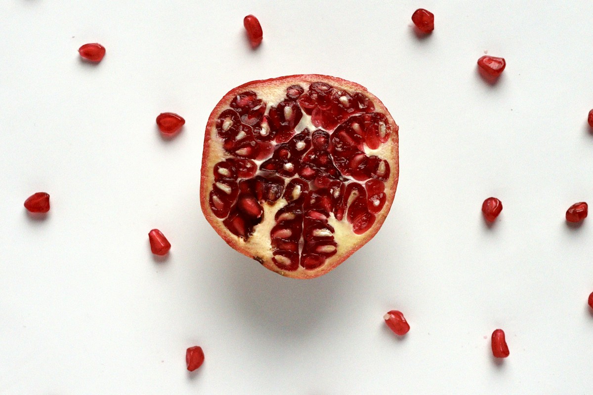
Dietary polyphenols represent the most abundant, structural diverse class of secondary plant metabolites in the human diet, encompassing over 8,000 identified compounds naturally concentrated in fruits, vegetables, green tea, cocoa, berries, nuts, and medicinal botanicals. Historically celebrated in nutritional science as simple direct free-radical scavengers in test-tube chemical assays, modern molecular pharmacology and pharmacokinetics have shattered this simplistic in vitro paradigm. In human physiology, parent polyphenols typically exhibit extraordinarily poor oral bioavailability: their large molecular weights, hydrophobic planar structures, and complex glycosidic linkages prevent significant absorption in the upper gastrointestinal tract, with over 90 to 95 percent traversing the small intestine intact to reach the dense microbial reactor of the human colon.
Within the colonic lumen, polyphenols encounter an ecological consortium of trillions of anaerobic commensal microorganisms expressing specialized catalytic enzymes that host human genomes lack – including beta-glucosidases, esterases, dehydroxylases, demethylases, and reductases. Through this process of microbial biotransformation, bulky, unabsorbable parent polyphenols are catabolized into a spectrum of low-molecular-weight, highly bioavailable bioactive postbiotic metabolites. The premier paradigm of this mutualistic co-metabolism is the enzymatic conversion of dietary ellagitannins and ellagic acid (concentrated in pomegranates, walnuts, and strawberries) into Urolithins, most prominently Urolithin A. Operating both locally in the gut mucosa and systemically following vascular absorption and hepatic phase II glucuronidation, Urolithin A functions as a potent biological signaling metabolite that crosses cellular membranes to stimulate selective mitochondrial autophagy (mitophagy), enhance vascular endothelial autophagy, and preserve neuromuscular vitality during human aging.
This comprehensive clinical nutrition and translational geroscience treatise explores the biochemical mechanisms, microbiome genetics, and systemic pharmacology of dietary polyphenol biotransformation. We analyze the chemical taxonomy of polyphenolic subclasses, dissect the multi-step enzymatic pathways of microbial catabolism, evaluate the pharmacokinetics and clinical stratification of human metabotypes (producers vs non-producers), examine Urolithin A-mediated PINK1-Parkin and SIRT1-AMPK signaling cascades, and review landmark human randomized clinical trials demonstrating improved muscular endurance, reduced systemic vascular stiffness, and attenuated biological senescence.
Chemical Taxonomy and Structural Classes of Dietary Polyphenols
Polyphenols are defined chemically as organic compounds possessing multiple phenolic hydroxyl groups attached to one or more aromatic benzene rings.
Dietary polyphenols are categorized into four major structural classes based on the number of phenol rings and the structural elements binding these rings together: flavonoids, phenolic acids, stilbenes, and lignans.
Flavonoids represent the largest and most widely distributed subclass, comprising a common diphenylpropane C6-C3-C6 carbon skeleton. Flavonoids are further subdivided into six principal subclasses: flavonols (quercetin, kaempferol in onions and apples), flavones (luteolin, apigenin in celery and parsley), flavan-3-ols (epicatechin, epigallocatechin gallate [EGCG] in green tea and cocoa), flavanones (hesperetin, naringenin in citrus fruits), anthocyanins (cyanidin, malvidin in dark berries), and isoflavones (daidzein, genistein in legumes and soybeans).
Phenolic acids comprise hydroxybenzoic acids (gallic acid, ellagic acid) and hydroxycinnamic acids (caffeic acid, ferulic acid, chlorogenic acid). Hydroxycinnamic acids are abundant in coffee, whole grains, and stone fruits, frequently conjugated to quinic acid or plant cell-wall polysaccharides.
Stilbenes, characterized by a 1,2-diphenylethylene carbon backbone, are exemplified by resveratrol, found in the skins of red grapes and Polygonum cuspidatum. Lignans, possessing a 2,3-dibenzylbutane structure, are concentrated in flaxseeds and sesame seeds.
In botanical tissues, polyphenols do not exist predominantly as free aglycones; rather, they are conjugated to sugars (glucose, rhamnose, galactose) as O-glycosides or C-glycosides, or polymerized into massive macromolecular complexes such as condensed tannins (proanthocyanidins) and hydrolyzable ellagitannins.
The Bioavailability Paradox: The Upper Gastrointestinal Absorption Barrier
For decades, the health-promoting benefits of polyphenol-rich diets were attributed to direct antioxidant scavenging within the bloodstream; however, rigorous pharmacokinetic measurements revealed a profound ‘bioavailability paradox’.
Parent dietary polyphenols exhibit dismal oral bioavailability: peak plasma concentrations (Cmax) of intact parent molecules rarely exceed low nanomolar thresholds (< 0.1 to 0.5 microM) following typical dietary intake, levels far below the concentrations required for direct chemical antioxidant scavenging in vivo.
This absorption barrier is dictated by physical chemistry and gastrointestinal physiology. Polyphenol glycosides are hydrophilic, bulky, and planar, preventing passive transcellular diffusion across the lipophilic apical membrane of small intestinal enterocytes.
While a minor fraction of specific monoglucosides (such as quercetin-4′-O-glucoside) can be hydrolyzed by the brush-border enzyme Lactase-Phlorizin Hydrolase (LPH) or transported via Sodium-Glucose Cotransporter 1 (SGLT1) followed by cytosolic beta-glucosidase (CBG) cleavage, over 90 to 95 percent of ingested polyphenols pass completely unabsorbed through the duodenum and jejunum.
Furthermore, any minor fraction absorbed into enterocytes is immediately subjected to intense Phase II conjugation – glucuronidation via UDP-glucuronosyltransferases (UGTs), sulfation via sulfotransferases (SULTs), and methylation via catechol-O-methyltransferase (COMT) – or pumped back into the intestinal lumen by ATP-binding cassette efflux transporters (P-glycoprotein / ABCB1 and BCRP / ABCG2).
The Colonic Microbiome as an Endocrine Metabolic Reactor
Because the small intestine is incapable of absorbing complex polyphenolic polymers and glycosides, the vast majority of dietary polyphenols reach the ileocecal valve and enter the dense, anaerobic ecosystem of the colon.
The human colon contains an extraordinary microbial density of over 10^11 to 10^12 bacterial cells per gram of luminal content, encoding an immense enzymatic repertoire that dwarfs human metabolic capabilities.
In the colon, dietary polyphenols encounter anaerobic commensals that utilize specialized carbohydrate-active enzymes (CAZymes), tannases, and aromatic ring-cleaving enzymes to metabolize polyphenols as auxiliary energetic substrates.
This mutualistic co-metabolic relationship operates bidirectionally: the gut microbiota breaks down polyphenols into small, absorbable bioactive metabolites, while polyphenols and their metabolites act as prebiotics, selectively inhibiting opportunistic pathobionts while stimulating the proliferation of beneficial commensals.
Through this microbial reactor, unabsorbable plant secondary metabolites are converted into potent, systemically bioavailable postbiotic signaling factors that achieve high micromolar concentrations in systemic circulation.
Microbial Catabolic Cascades: Deglycosylation, Dehydroxylation, and Ring Fission
The microbial biotransformation of dietary polyphenols follows a conserved, sequential series of biochemical reactions executed by specialized bacterial consortia.
Step 1: Deglycosylation. Colonic anaerobes express diverse glycosyl hydrolases – including alpha-L-rhamnosidases, beta-D-glucosidases, and beta-D-galactosidases – that cleave sugar moieties from the polyphenol aglycone, liberating free hydrophobic phenolic aglycones.
Step 2: C-Ring Fission and Hydrolysis. Following deglycosylation, bacterial enzymes execute carbon-carbon bond cleavage and ring-opening reactions. In flavonoids, bacterial chalcone isomerases and reductases cleave the central heterocyclic C-ring, breaking the diphenylpropane skeleton into smaller phenolic fragments.
Step 3: Dehydroxylation and Demethylation. Aromatic dehydroxylases selectively remove hydroxyl (-OH) groups from specific positions on the benzene rings, while demethylases remove methyl ether groups, generating simplified phenolic metabolites.
Step 4: Alpha- and Beta-Oxidation. The resulting aliphatic side chains undergo sequential microbial oxidation and reduction, converting complex botanical structures into simple, bioavailable low-molecular-weight phenolic acids: phenylpropionic acids, phenylacetic acids, and hydroxybenzoic acids.
These catabolic intermediates are easily absorbed across the colonic mucosal epithelium via passive non-ionic diffusion and monocarboxylate transporters (MCTs), entering the mesenteric venules to reach the liver and systemic circulation.
Ellagitannins and Ellagic Acid: The Botanical Precursors of Urolithins
Among all dietary polyphenolic classes, the hydrolyzable ellagitannins provide the most compelling illustration of clinical microbiome-dependent bioactivation.
Ellagitannins are complex, high-molecular-weight polymers composed of hexahydroxydiphenic acid (HHDP) units esterified to a central D-glucose core. Premier dietary sources include the pomegranate (Punica granatum, rich in punicalagins A and B), walnuts (Juglans regia, rich in pedunculagin), black raspberries, strawberries, and oak-aged wines (rich in castalagin and vescalagin).
Punicalagin, the major ellagitannin of pomegranate husk and arils, is a massive molecule with a molecular weight of 1,084 Daltons, possessing zero oral bioavailability in the upper digestive tract.
Upon reaching the distal small intestine and colon, punicalagins are hydrolyzed by microbial esterases and intestinal pH shifts into monomeric ellagic acid (a planar dilactone of hexahydroxydiphenic acid, MW 302 Da).
While ellagic acid is smaller, its planar, symmetrical four-ring structure renders it extraordinarily insoluble in aqueous physiological fluids, severely limiting its systemic absorption and ensuring its passage into the colonic microbiome for downstream biotransformation.
The Urolithin Biosynthetic Pathway: Stepwise Microbial Lactone Cleavage
In the colonic lumen, ellagic acid undergoes a multi-step enzymatic biotransformation cascade executed by specialized anaerobic bacteria to synthesize the family of Urolithins.
Urolithins are dibenzo[b,d]pyran-6-one derivatives characterized by varying numbers and positions of phenolic hydroxyl groups. The catabolic sequence begins with the opening and decarboxylation of one of the two lactone rings of ellagic acid, yielding pentahydroxy-urolithin (Urolithin M-5).
Sequential dehydroxylation reactions follow in precise chemical order: Urolithin M-5 is converted into tetrahydroxy-urolithin (Urolithin M-6), which is subsequently dehydroxylated to trihydroxy-urolithin (Urolithin C).
Urolithin C undergoes further bacterial dehydroxylation to yield dihydroxy-urolithins: Urolithin A (3,8-dihydroxy-urolithin) and Urolithin B (3-hydroxy-urolithin).
Under specific microbial conditions, Urolithin A can be terminal dehydroxylated into monohydroxy-urolithin B or isourolithin A (3,9-dihydroxy-urolithin). Because Urolithins possess a much smaller molecular weight (~228 Da) and significantly higher lipophilicity than ellagic acid, they are readily absorbed across the colonic mucosal epithelium into the portal venous circulation.
Identification of the Producer Microorganisms: Gordonibacter and Clostridium
A major scientific milestone in microbiome biochemistry was the isolation and genomic characterization of the specific bacterial species responsible for executing urolithin synthesis.
In 2014, David Beltran and colleagues isolated Gordonibacter urolithinfaciens and Gordonibacter pamelaeae – novel Gram-positive, strictly anaerobic asporogenous coccobacilli belonging to the Coriobacteriaceae family within the Actinomycetota phylum.
Functional enzymatic assays confirmed that Gordonibacter strains catalyze the initial steps of the cascade, converting ellagic acid into Urolithin M-5, Urolithin M-6, and Urolithin C; however, pure cultures of Gordonibacter cannot perform the final dehydroxylation steps required to produce Urolithin A.
In 2020, researchers isolated Ellagibacter isourolithinifaciens, which converts Urolithin C into isourolithin A, alongside specific strains belonging to the Clostridiales order (including Ruminococcaceae species) that express the terminal dehydroxylases executing conversion to Urolithin A.
This modular metabolic division of labor proves that urolithin production requires a functional microbial cross-feeding consortium rather than a single bacterial species operating in isolation.
The Human Urolithin Metabotypes: Phenotype A, Phenotype B, and Non-Producers
An extraordinary clinical discovery pioneered by Francisco Tomas-Barberan and Juan Carlos Espin is that human populations exhibit marked inter-individual stratification in their ability to produce urolithins, a phenomenon designated ‘human urolithin metabotypes’.
Following standardized dietary intake of ellagitannins or pomegranate juice, human individuals stratify into three distinct, stable metabotypes based on their urinary and plasma urolithin excretion profiles:
Metabotype A (Phenotype A): Individuals who exclusively produce Urolithin A conjugates, lacking isourolithin A or Urolithin B. This phenotype comprises approximately 40 to 60 percent of healthy young populations and is associated with optimal colonic microbial diversity and favorable cardiometabolic biomarkers.
Metabotype B (Phenotype B): Individuals who produce Urolithin B and isourolithin A in addition to Urolithin A. Comprising 20 to 30 percent of populations, Phenotype B is significantly more prevalent in older adults, individuals with obesity, metabolic syndrome, and inflammatory bowel diseases, serving as a clinical biomarker of intestinal dysbiosis.
Metabotype 0 (Non-Producers): Individuals who completely fail to produce any urolithins (producing zero Urolithin A, B, or C). Comprising 10 to 20 percent of humans, non-producers completely lack the specific Coriobacteriaceae and Clostridial taxa required for ellagic acid biotransformation, deriving zero systemic urolithin benefits from consuming pomegranate or walnuts.
This stratification provides a biological explanation for the conflicting results of historical dietary trials and underscores the clinical necessity of direct, standardized postbiotic Urolithin A supplementation.
Hepatic Phase II Metabolism and Systemic Pharmacokinetics of Urolithin A
Following colonic absorption, unconjugated Urolithin A enters the portal venous circulation and is transported directly to the liver.
In hepatocytes, Urolithin A is rapidly recognized as a xenobiotic compound and subjected to extensive Phase II hepatic conjugation.
The primary circulating human metabolite is Urolithin A glucuronide (Urolithin A-3-O-glucuronide), synthesized via UDP-glucuronosyltransferase 1A3 (UGT1A3) and UGT1A8, alongside minor proportions of Urolithin A sulfate.
Pharmacokinetic investigations in healthy human volunteers demonstrate that oral Urolithin A administration yields rapid, dose-dependent plasma absorption, reaching peak plasma concentrations (Cmax) within 4 to 8 hours with an elimination half-life (t1/2) of approximately 17 to 20 hours, supporting convenient once-daily clinical dosing.
Crucially, in target peripheral tissues experiencing localized inflammation, cellular stress, or energetic demand, tissue beta-glucuronidase enzymes cleave the glucuronide moiety, locally liberating free, lipophilic unconjugated Urolithin A to execute its intracellular signaling actions.
Urolithin A and Mitophagy: The Molecular Mechanism of Mitochondrial Rejuvenation
The premier biological discovery that established Urolithin A as a breakthrough geroscience therapeutic was published in 2016 by Johan Auwerx, Patrick Aebischer, and colleagues in Nature Medicine.
Auwerx demonstrated that Urolithin A is a potent, selective inducer of mitophagy – the specialized form of macroautophagy that identifies, engulfs, and lysosomally degrades damaged, dysfunctional, and depolarized mitochondria.
Mechanistically, Urolithin A induces a mild, transient, sub-lethal decrease in mitochondrial membrane potential without causing cellular toxicity or ATP depletion. This slight depolarization stabilizes PTEN-Induced Kinase 1 (PINK1) on the outer mitochondrial membrane.
Accumulated PINK1 phosphorylates ubiquitin at Ser65, which recruits and allosterically activates the cytosolic E3 ubiquitin ligase Parkin (PRKN). Activated Parkin polyubiquitinates outer mitochondrial membrane proteins (MFN1, MFN2, VDAC1), generating dense phospho-ubiquitin coats that recruit autophagy receptor adaptors (p62/SQSTM1, Optineurin).
These adaptors bridge the damaged organelle to lipidated LC3-II on growing phagophore membranes, encapsulating the defective mitochondrion within an autophagosome for enzymatic lysosomal clearance, stimulating healthy mitochondrial biogenesis to replace degraded organelles.
SIRT1 and AMPK Signaling: Coordinating Bioenergetic Flexibility
In parallel with the PINK1-Parkin cascade, Urolithin A activates the master metabolic energy sensors of mammalian cells: Sirtuin-1 (SIRT1) and AMP-activated Protein Kinase (AMPK).
By stimulating mitophagy, Urolithin A improves cellular respiratory efficiency and optimizes the intracellular NAD+:NADH and AMP:ATP ratios. Elevated NAD+ activates the nuclear and mitochondrial deacetylase SIRT1.
Active SIRT1 deacetylates Peroxisome Proliferator-Activated Receptor Gamma Coactivator 1-alpha (PGC-1alpha) at multiple lysine residues, driving PGC-1alpha into its transcriptionally active conformation.
Concurrently, active AMPK phosphorylates PGC-1alpha at Thr177 and Ser538. Synergistically activated PGC-1alpha translocates to the nucleus, coactivating Nuclear Respiratory Factors 1 and 2 (NRF-1, NRF-2) and Mitochondrial Transcription Factor A (TFAM).
This transcriptional program stimulates de novo mitochondrial DNA replication, structural cristae biogenesis, and electron transport chain complex synthesis, coupling the clearance of damaged organelles to the rapid generation of youthful, high-capacity respiratory networks.
Endothelial Autophagy and Vascular Health: Arterial Stiffness Reversal
Vascular endothelial dysfunction and progressive arterial stiffening represent foundational hallmarks of human cardiovascular aging, predisposing individuals to isolated systolic hypertension, coronary artery disease, and heart failure.
The vascular endothelium relies strictly on baseline basal autophagy to clear oxidized macromolecules, preserve endothelial nitric oxide synthase (eNOS) coupling, and maintain cellular mechanical elasticity.
In aging blood vessels, endothelial autophagy declines sharply, resulting in the intracellular accumulation of protein aggregates, reactive oxygen species, and inflammatory mediators.
Urolithin A readily penetrates vascular endothelial cells, where it potently stimulates endothelial autophagy via the inhibition of mTORC1 and activation of ULK1.
Restored endothelial autophagy preserves the eNOS cofactor tetrahydrobiopterin (BH4), preventing eNOS uncoupling and dramatically enhancing flow-mediated vasodilation (FMD). In preclinical and clinical trials, Urolithin A administration significantly attenuated aortic pulse wave velocity (PWV), reversed arterial stiffness, and suppressed vascular cell adhesion molecule 1 (VCAM-1) expression.
Anti-Inflammatory Modulation: Suppressing NF-kB and the NLRP3 Inflammasome
Beyond organellar quality control, Urolithin A acts as a powerful anti-inflammatory postbiotic metabolite, suppressing both systemic and localized sterile tissue inflammation.
In monocytes, macrophages, and microglial cells, Urolithin A inhibits the phosphorylation of IkappaB kinase (IKK), preventing the degradation of IkappaB-alpha and blocking the nuclear translocation of the p65/p50 heterodimer of Nuclear Factor kappa B (NF-kB).
Shutting down NF-kB transcription potently represses the synthesis of core pro-inflammatory cytokines, including Tumor Necrosis Factor-alpha (TNF-alpha), Interleukin-6 (IL-6), and Interleukin-1beta (IL-1beta).
Furthermore, Urolithin A directly suppresses the assembly of the NLRP3 inflammasome by inhibiting mitochondrial reactive oxygen species leakage and preventing the release of oxidized mitochondrial DNA into the cytosol.
In animal models of colitis, arthritis, and atherosclerosis, oral Urolithin A significantly reduced circulating high-sensitivity C-reactive protein (hs-CRP) and mucosal inflammatory infiltrates, proving that mitophagy induction cleanly neutralizes upstream triggers of chronic sterile inflammation.
Human Clinical Trials: Muscle Endurance and Cellular Healthspan
Urolithin A has successfully crossed the translational divide into human clinical medicine, with multiple rigorous, double-blind, randomized, placebo-controlled clinical trials validating its efficacy and safety.
In a landmark Phase I/II clinical trial published in Nature Metabolism in 2019, David Ryu, Johan Auwerx, and colleagues evaluated oral Urolithin A (Mitopure, 250 mg, 500 mg, and 1,000 mg daily for 4 weeks) in sedentary elderly adults (ages 61 to 85).
The trial met all primary safety endpoints, demonstrating excellent oral tolerability without adverse events. Molecular analyses of skeletal muscle biopsies revealed that Urolithin A significantly upregulated mitochondrial gene expression signatures, stimulated muscle mitophagy, and increased plasma acylcarnitines, reflecting improved fatty acid beta-oxidation.
In 2022, a pivotal randomized clinical trial published in JAMA Network Open evaluated daily oral Urolithin A (1,000 mg) for four months in older adults. Participants receiving Urolithin A experienced significant, clinically meaningful improvements in 6-minute walk distance (increasing by an average of 33.5 meters over placebo) and marked improvements in muscle endurance (measured by handgrip and leg muscle contraction cycles to fatigue).
Furthermore, circulating plasma biomarkers of systemic inflammation, including sTNFR1 and pro-inflammatory acylcarnitines, declined significantly, establishing Urolithin A as the first clinically proven nutritional postbiotic to enhance human muscle performance via mitophagy.
Cardiometabolic Syndrome: Lipid Profiles and Hepatic Steatosis Attenuation
Metabolic syndrome – characterized by visceral adiposity, hypertriglyceridemia, low HDL cholesterol, insulin resistance, and hepatic steatosis – is driven by cellular mitochondrial overload and incomplete fatty acid oxidation.
In preclinical models of diet-induced obesity and non-alcoholic fatty liver disease (MASLD), oral administration of Urolithin A produced dramatic metabolic improvements: reducing body weight gain, improving whole-body insulin sensitivity, and substantially attenuating intrahepatic triglyceride accumulation.
Mechanistically, Urolithin A stimulates thermogenic brown adipose tissue (BAT) activation and induces ‘browning’ of subcutaneous white adipose tissue (beige adipocyte differentiation), significantly increasing uncoupling protein 1 (UCP1) expression and non-shivering thermogenesis.
In the liver, Urolithin A enhances hepatic mitochondrial fatty acid oxidation while suppressing Sterol Regulatory Element-Binding Protein 1c (SREBP-1c) and Fatty Acid Synthase (FASN), shutting down hepatic de novo lipogenesis.
Clinical lipid profiles in human trial participants demonstrate significant reductions in plasma triglycerides and increases in the cardioprotective HDL-to-triglyceride ratio, confirming broad cardiometabolic utility.
Neuroprotection: Blood-Brain Barrier Penetration and Microglial Restabilization
As an uncharged, lipophilic low-molecular-weight compound, unconjugated Urolithin A possesses the biophysical capacity to cross the blood-brain barrier via passive transcellular diffusion, achieving detectable concentrations in the central nervous system parenchyma.
Within the brain, neurons and astrocytes maintain immense baseline bioenergetic demands, rendering them exceptionally vulnerable to the accumulation of damaged mitochondria and oxidative stress.
In preclinical models of Alzheimer’s disease (including APP/PS1 and 3xTg-AD transgenic mice), Urolithin A administration significantly reduced cerebral amyloid-beta (Abeta) plaque burdens, attenuated tau hyperphosphorylation, and reversed spatial learning and memory deficits on the Morris water maze.
Mechanistically, Urolithin A stimulates mitophagy within cortical neurons, restoring mitochondrial ATP synthesis and preventing the bioenergetic collapse that drives synaptic retraction.
Concurrently, Urolithin A suppresses microglial neuroinflammation, shutting down the release of neurotoxic nitric oxide and IL-1beta, directly preserving hippocampal dentate gyrus adult neurogenesis and synaptic plasticity.
Dermal Extracellular Matrix Preservation: Type I Collagen and Photoprotection
Human cutaneous aging is characterized by progressive fragmentation of dermal extracellular matrix scaffolds, loss of structural type I collagen, and impaired microvascular perfusion, accelerated by solar ultraviolet (UV) radiation.
Exposure of human dermal fibroblasts to UV-A and UV-B radiation generates massive bursts of reactive oxygen species, triggering mitochondrial DNA deletions, respiratory chain collapse, and the upregulation of matrix metalloproteinase-1 (MMP-1 / interstitial collagenase).
Active MMP-1 enzymatically degrades triple-helical type I and type III collagen fibers, leading to deep cutaneous wrinkling, dermal thinning, and loss of mechanical elasticity.
Urolithin A penetrates dermal fibroblasts, where it potently stimulates mitochondrial quality control, suppresses UV-induced ROS leakage, and blocks NF-kB and AP-1 transcription factor activation.
Consequently, Urolithin A significantly inhibits MMP-1 and MMP-3 expression while upregulating procollagen type I C-peptide synthesis, preserving extracellular matrix architecture and providing profound systemic and topical photoprotection against extrinsic photoaging.
Other Microbial Postbiotics: Equol, Enterolactone, and 3-HPAA
While Urolithin A represents the premier paradigm of polyphenol biotransformation, the colonic microbiome generates a diverse universe of bioactive postbiotic metabolites from other botanical polyphenol classes.
Equol: Daidzein, a major isoflavone found in soybeans, is biotransformed by specialized colonic anaerobes (such as Adlercreutzia equolifaciens and Slackia isoflavoniconvertens) into Equol (7-hydroxy-3-(4′-hydroxyphenyl)chroman). Equol exhibits significantly higher affinity for estrogen receptor beta (ER-beta) and much higher antioxidant potency than parent daidzein, conferring cardiovascular protection and relieving menopausal symptoms in ‘equol-producing’ individuals.
Enterolactone and Enterodiol: Dietary lignans (secoisolariciresinol, matairesinol) from flaxseed are metabolized by Eggerthella lenta and Blautia producta into the mammalian enterolignans enterodiol and enterolactone, which exert weak estrogenic, anti-angiogenic, and anti-proliferative effects against breast and prostate cancers.
3-Hydroxyphenylacetic Acid (3-HPAA): Proanthocyanidins from grape seeds and cocoa are fermented into 3-HPAA and 3,4-dihydroxyphenylpropionic acid, which penetrate vascular smooth muscle to stimulate nitric oxide release and lower systemic blood pressure.
These diverse postbiotics prove that the human microbiome functions as an indispensable biological biofactory, translating dietary plant inputs into systemic hormonal and metabolic regulators.
Dietary Optimization: Cultivating an Optimal Polyphenol-Metabolizing Microbiome
Given that 10 to 20 percent of humans are non-producers (Metabotype 0) and 20 to 30 percent harbor the dysbiotic Phenotype B, a vital clinical question is whether individuals can cultivate a high-capacity polyphenol-metabolizing microbiome through lifestyle and nutrition.
Microbial alpha-diversity is the single most decisive predictor of polyphenol biotransformation capacity: individuals consuming high dietary fiber diversity (exceeding 30 distinct plant varieties per week) exhibit significantly higher abundances of Coriobacteriaceae and Clostridial producer species.
Consuming prebiotic fibers – particularly resistant starch, inulin, and beta-glucans – stimulates the proliferation of primary fermenters that produce short-chain fatty acids (acetate, butyrate), creating an optimal acidic colonic luminal pH (pH 5.5 to 6.5) that favors Gordonibacter enzymatic activity.
Furthermore, regular, sustained consumption of diverse polyphenol-rich whole foods (the Mediterranean-DASH diet) exerts a prebiotic-like selective pressure: polyphenols suppress pathogenic Clostridium perfringens while providing a continuous substrate that maintains the abundance of polyphenol-degrading bacterial consortia.
Avoiding repeated unnecessary broad-spectrum antibiotic courses and minimizing ultra-processed dietary emulsifiers prevents the catastrophic eradication of delicate Coriobacteriaceae and Ruminococcaceae lineages.
Clinical Supplementation Protocols: Purified Postbiotic Urolithin A
While dietary optimization with pomegranates, walnuts, and berries is universally recommended for general health, relying solely on whole foods cannot guarantee therapeutic Urolithin A bioavailability due to widespread inter-individual metabotype differences.
Clinical pharmacokinetic studies demonstrate that drinking 500 mL of 100% pomegranate juice daily fails to generate detectable circulating Urolithin A in over 60 percent of individuals (Metabotype 0 non-producers and poor converters), while delivering over 60 grams of liquid fruit sugars.
To overcome this biological limitation, medical geroscience developed standardized, highly purified synthetic or microbially fermented Urolithin A (such as Mitopure) for direct oral administration.
Direct oral Urolithin A administration completely bypasses the gut microbiome bottleneck: 100 percent of individuals achieve therapeutic, micromolar circulating plasma concentrations of Urolithin A and its active conjugates, regardless of baseline microbiome composition or metabotype status.
Clinical dosing guidelines derived from published trials recommend an oral dosage of 500 mg to 1,000 mg daily taken with or without food, providing consistent, reliable stimulation of systemic mitophagy and muscle performance.
Future Horizons: Metagenomic Engineering and Synthetic Postbiotic Consortia
The clinical frontier of polyphenol pharmacology is advancing from empirical nutritional advice toward precision microbiome engineering and synthetic biology.
High-throughput shotgun metagenomic sequencing and functional CAZyme profiling will soon allow clinicians to rapidly analyze a patient’s stool sample to determine their precise polyphenol-metabolizing enzymatic capacity, identifying specific missing catabolic steps.
Pharmaceutical research is developing defined synthetic bacterial consortia – combining Gordonibacter urolithinfaciens, Ellagibacter isourolithinifaciens, and specialized Clostridiales strains into an oral, enterically coated biotherapeutic capsule.
Administering these synthetic consortia alongside standardized ellagitannin prebiotics will permanently convert non-producing Metabotype 0 individuals into high-efficiency Urolithin A producers.
By fusing microbiology, synthetic biology, and molecular pharmacology, medicine will harness the full therapeutic power of the botanical kingdom to expand human vitality, decelerate biological aging, and promote lifelong healthspan.
| Parent Botanical Polyphenol | Primary Dietary Sources | Colonic Microbial Metabolite (Postbiotic) | Key Producer Microbial Species | Validated Cellular & Clinical Efficacy |
|---|---|---|---|---|
| Ellagitannins / Ellagic Acid | Pomegranates, walnuts, strawberries, oak-aged wine | Urolithin A (3,8-dihydroxy-urolithin) | Gordonibacter urolithinfaciens, Clostridiales | Stimulates PINK1-Parkin mitophagy; increases muscle endurance & 6MWD |
| Soy Isoflavones (Daidzein) | Soybeans, tofu, tempeh, legumes | Equol (7-hydroxy-3-(4-hydroxyphenyl)chroman) | Adlercreutzia equolifaciens, Slackia isoflavoniconvertens | High-affinity ER-beta binding; vasodilation & bone mineral density retention |
| Dietary Lignans (Secoisolariciresinol) | Flaxseeds, sesame seeds, rye whole grains | Enterolactone & Enterodiol (Mammalian Lignans) | Eggerthella lenta, Blautia producta | Anti-angiogenic; lower cardiovascular mortality & hormone-sensitive cancer risk |
| Proanthocyanidins / Flavan-3-ols | Cocoa, green tea, dark berries, grape seeds | 5-(3′,4′-dihydroxyphenyl)-gamma-valerolactone | Flavonifractor plautii | Restores endothelial eNOS coupling; lowers central blood pressure |
| Flavonols (Quercetin Glycosides) | Red onions, capers, apples, kale | 3,4-Dihydroxyphenylacetic acid (3,4-DHPAA) | Eubacterium ramulus | Inhibits vascular smooth muscle proliferation & platelet hyper-aggregation |
The comparative matrix above illustrates the profound transformation that occurs when parent dietary polyphenols undergo colonic microbial biotransformation into bioavailable postbiotic metabolites. By examining the specific botanical precursors, key microbial species, resulting postbiotic compounds, and validated clinical endpoints across major polyphenol subclasses, clinicians can understand how the gut microbiome acts as an essential metabolic partner.
Recognizing the absolute dependency of polyphenol efficacy upon specific microbial consortia highlights why dietary interventions produce variable clinical outcomes across diverse patient populations, establishing the clinical rationale for precision microbiome diagnostics and standardized postbiotic supplementation.
Frequently Asked Questions Regarding Polyphenol Metabolism and Urolithin A
Why are parent dietary polyphenols so poorly absorbed in the human digestive tract?
Parent dietary polyphenols exhibit poor oral bioavailability (typically less than 5 to 10 percent) because they are large, complex molecules naturally bound to sugars (glycosides) or polymerized into dense macromolecules (such as tannins). Their high hydrophilicity and bulky planar structures prevent them from crossing the lipophilic cell membranes of small intestinal enterocytes. As a result, over 90 percent pass completely unabsorbed into the colon, where they must be metabolized by gut bacteria.
What is Urolithin A and how is it produced in the human body?
Urolithin A is a natural postbiotic metabolite produced by the gut microbiome from dietary ellagitannins and ellagic acid, compounds found in pomegranates, walnuts, and berries. In the colon, specialized anaerobic bacteria (including Gordonibacter urolithinfaciens and Clostridial species) perform sequential ring-cleavage and dehydroxylation reactions, converting ellagic acid into Urolithin A, which is readily absorbed into the bloodstream.
What are the human urolithin metabotypes and why do they matter clinically?
Humans stratify into three distinct urolithin metabotypes based on their gut microbiome composition: Metabotype A (produce only Urolithin A, associated with optimal health), Metabotype B (produce Urolithin B and isourolithin A, associated with dysbiosis and metabolic disease), and Metabotype 0 (non-producers who produce zero urolithins). Approximately 10 to 20 percent of humans are non-producers who derive no urolithin benefits from eating pomegranates or walnuts, making direct postbiotic supplementation necessary.
How does Urolithin A stimulate mitophagy in human cells?
Urolithin A stimulates mitophagy (the selective removal of damaged mitochondria) by inducing a mild, physiological decrease in mitochondrial membrane potential. This stabilizes PINK1 kinase on the outer mitochondrial membrane, which recruits and activates the E3 ubiquitin ligase Parkin. Parkin tags the damaged organelle with ubiquitin chains, signaling autophagosomes to engulf the defective mitochondrion and deliver it to lysosomes for destruction, stimulating healthy mitochondrial renewal.
What clinical evidence supports Urolithin A for muscle endurance?
In multiple randomized, double-blind, placebo-controlled clinical trials published in Nature Metabolism (2019) and JAMA Network Open (2022), daily oral administration of Urolithin A (500 to 1,000 mg) in middle-aged and older adults significantly increased 6-minute walk distance, improved muscle endurance during repetitive contraction tests, and decreased circulating inflammatory markers (sTNFR1), without requiring changes in baseline exercise.
Can drinking pomegranate juice replace direct Urolithin A supplementation?
No, drinking pomegranate juice cannot reliably replace direct Urolithin A supplementation for two reasons: first, over 60 percent of individuals are either non-producers or poor converters who lack the specific colonic bacteria required to synthesize Urolithin A; second, drinking sufficient pomegranate juice to attempt therapeutic conversion delivers high amounts of liquid fruit sugars (over 60 grams per half-liter), which can elevate blood glucose and drive hepatic de novo lipogenesis.
How does Urolithin A improve vascular health and arterial stiffness?
Urolithin A penetrates vascular endothelial cells and activates endothelial autophagy via SIRT1-AMPK signaling. Restored autophagy clears damaged cellular debris and uncoupled endothelial nitric oxide synthase (eNOS), increasing the bioavailability of vasoprotective nitric oxide (NO). This enhances flow-mediated vasodilation, reduces aortic pulse wave velocity (arterial stiffness), and suppresses inflammatory adhesion molecules (VCAM-1).
What other beneficial postbiotic metabolites are produced from polyphenols?
Beyond Urolithin A, the gut microbiota produces several vital postbiotics: Equol (produced from soy daidzein by Adlercreutzia equolifaciens, providing estrogenic and cardioprotective benefits), Enterolactone and Enterodiol (produced from flaxseed lignans by Eggerthella lenta, protecting against hormone-sensitive cancers), and phenylvalerolactones (produced from cocoa and green tea flavan-3-ols, lowering blood pressure).
How can an individual improve their gut microbiome’s ability to metabolize polyphenols?
An individual can optimize their polyphenol-metabolizing capacity by increasing dietary plant diversity (aiming for over 30 distinct whole plant foods per week), consuming diverse prebiotic fermentable fibers (inulin, resistant starch, beta-glucans), and regularly eating polyphenol-rich foods that exert selective prebiotic pressure. Avoiding unnecessary broad-spectrum antibiotic exposure preserves the delicate anaerobic Coriobacteriaceae species required for biotransformation.
What is the recommended clinical dosage of purified Urolithin A?
Published human clinical trials have established that oral Urolithin A (Mitopure) is safe, well-tolerated, and effective at dosages between 500 mg and 1,000 mg once daily. It can be taken with or without food and produces predictable, micromolar circulating plasma concentrations in 100 percent of recipients, completely overcoming the gut microbiome metabotype bottleneck.
Clinical Summary and Translational Nutrition Horizons
The convergence of nutritional biochemistry, clinical gastroenterology, and geroscience has radically transformed our understanding of dietary polyphenols. The historical view that polyphenols function as simple direct free-radical scavengers in the bloodstream has been completely superseded by a nuanced ecological paradigm recognizing the colonic microbiome as an indispensable bio-transformative partner.
By traversing the small intestine unabsorbed, parent polyphenols reach the colonic anaerobic reactor, where specialized commensal consortia execute precision ring-fission, deglycosylation, and dehydroxylation cascades to synthesize bioavailable postbiotic signaling molecules. As exemplified by Urolithin A, these microbially derived postbiotics cross cellular membranes to stimulate selective mitochondrial autophagy (mitophagy), enhance endothelial autophagy, suppress sterile inflammation, and restore neuromuscular functional capacity during biological aging.
Yet, the discovery of human urolithin metabotypes reveals profound inter-individual disparities: significant portions of the global population lack the essential microbial machinery required to synthesize these longevity metabolites from whole foods alone. Bridging this gap through precision microbiome diagnostics, dietary prebiotic diversification, and standardized postbiotic Urolithin A supplementation represents a triumphant milestone in evidence-based metabolic and preventive medicine, empowering clinicians to optimize cellular energy and preserve human vitality across the lifespan.
For accredited institutional consensus guidelines, clinical trial registries, and educational resources regarding polyphenol metabolism and mitochondrial biology, clinicians are encouraged to consult the American Society for Nutrition (ASN), the National Institute on Aging (NIA), and the Nature Metabolism Clinical Forum. Peer-reviewed research literature is continuously indexed on PubMed National Library of Medicine, alongside global nutrition and healthy aging directives from the World Health Organization.
