


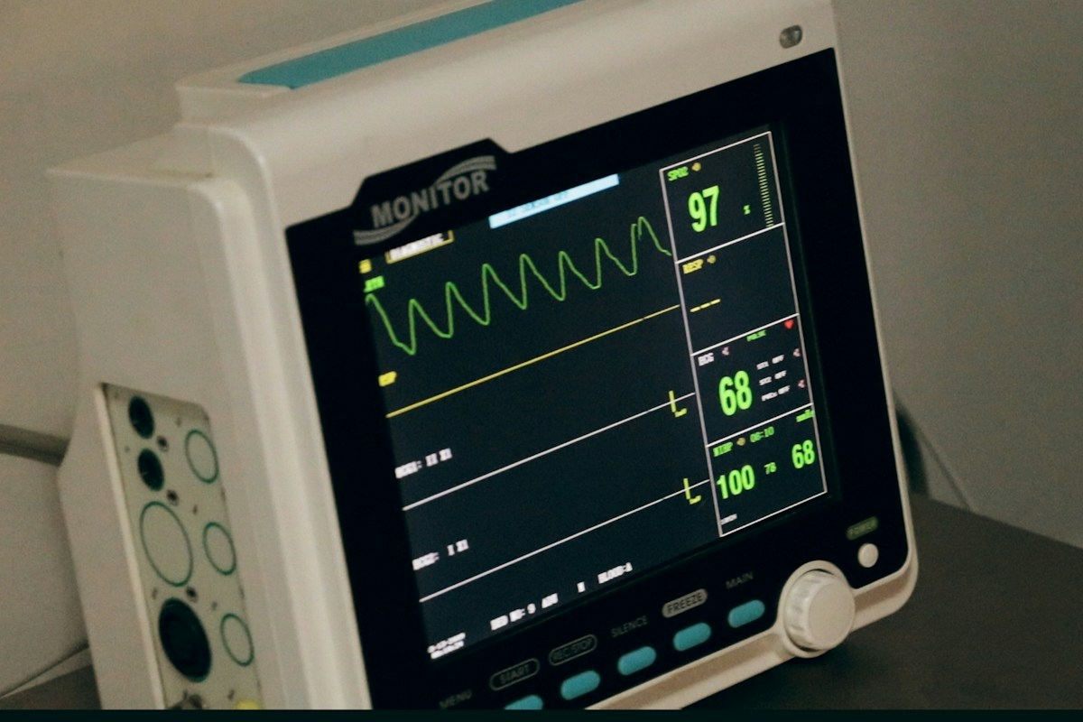
Heart rate variability (HRV) has emerged as one of the most vital non-invasive biomarkers in modern exercise physiology, clinical cardiology, and sports neurobiology, offering an objective window into the real-time regulatory balance of the human autonomic nervous system (ANS). Contrary to the intuitive assumption that a healthy resting heart beats with rigid metronomic precision, a healthy cardiovascular system displays profound beat-to-beat variability in the intervals between successive heartbeats (R-R intervals). This dynamic fluctuation reflects the constant push-pull interplay between the decelerating parasympathetic (vagal) branch and the accelerating sympathetic branch of the autonomic nervous system.
In high-performance endurance athletics, occupational stress management, and preventative cardiology, tracking longitudinal heart rate variability provides actionable insights into internal physiological load, allostatic stress accumulation, and systemic recovery status. A robust baseline level of heart rate variability indicates superior parasympathetic cardiac modulation, elevated cardiorespiratory fitness, heightened endothelial elasticity, and neurological resiliency against environmental stressors. Conversely, sustained suppression of vagally mediated HRV indices serves as an early clinical harbinger of non-functional overreaching, overtraining syndrome, systemic neuroendocrine exhaustion, or subclinical infectious illness.
This comprehensive clinical guide delivers an exhaustive exploration of heart rate variability physiology and methodology, detailing the cardiac neuroanatomy of sinoatrial node innervation, the mathematical derivations and physiological significance of time-domain (RMSSD, SDNN), frequency-domain (HF, LF), and non-linear indices, and the neurobiological interactions between nocturnal HRV and sleep architecture. Furthermore, we provide evidence-based clinical algorithms for establishing individualized rolling baselines, utilizing the Smallest Worthwhile Change (SWC) metric to guide athletic training periodization and safeguard long-term cardiorespiratory health.
Neuroanatomy and Electrophysiology: Sinoatrial Node Autonomic Regulation
Cardiac automaticity originates within the primary biological pacemaker of the heart, the sinoatrial (SA) node, situated subepicardially at the junction of the superior vena cava and the right atrium. While pacemaker cells within the sinoatrial node generate spontaneous action potentials via the hyperpolarization-activated cyclic nucleotide-gated (HCN4) channels (responsible for the inward ‘funny’ pacemaker current, If), the intrinsic firing frequency of approximately 100 beats per minute is continuously modulated by tonic autonomic neurochemical inputs.
Parasympathetic cardiac regulation is conveyed through the bilateral vagus nerves (Cranial Nerve X), whose preganglionic fibers originate within the nucleus ambiguus and the dorsal motor nucleus of the vagus inside the medulla oblongata. These myelinated axons travel down the carotid sheath into cardiac plexuses and synapse onto postganglionic parasympathetic ganglia embedded within epicardial fat pads adjacent to the sinoatrial node. Postganglionic terminals release acetylcholine (ACh), which binds to cardiac muscarinic type 2 (M2) receptors coupled to Gi/o inhibitory heterotrimeric G-proteins.
Activation of M2 receptors liberates beta-gamma G-protein subunits that directly bind to and open inward-rectifying potassium channels (GIRK1/4, also termed IKACh), hyperpolarizing the pacemaker cell membrane. Concurrently, the alpha-i subunit inhibits adenylate cyclase, reducing intracellular cyclic AMP (cAMP) concentrations and decreasing protein kinase A (PKA) activity. This biochemical cascade blunts L-type calcium channel (ICa,L) and funny current (If) conduction, flattening phase 4 diastolic depolarization and decelerating heart rate within 150 to 400 milliseconds. Because acetylcholine is rapidly cleared by abundant acetylcholinesterase in the sinoatrial interstitium, parasympathetic modulation occurs almost instantaneously, allowing beat-to-beat tracking of cardiovagal flux.
In stark contrast, sympathetic cardiac regulation operates via postganglionic unmyelinated fibers originating in the cervical and thoracic sympathetic chain ganglia (stellate ganglion, T1-T4). These nerve terminals release norepinephrine, which binds to cardiac beta-1 adrenergic receptors coupled to Gs stimulatory proteins. Activation of adenylate cyclase escalates cAMP production, activating PKA to phosphorylate L-type calcium channels, ryanodine receptors (RyR2), and phospholamban (PLN), steepening phase 4 depolarization and accelerating conduction velocity. However, because norepinephrine reuptake and secondary messenger phosphorylation require several seconds, sympathetic modulation is incapable of generating high-frequency, beat-to-beat fluctuations, functioning instead across slower, low-frequency physiological timescales.
Time-Domain HRV Metrics: Mathematical Derivations and Physiological Meaning
Time-domain metrics represent the most accessible, computationally robust, and clinically validated indices of heart rate variability, derived from the mathematical analysis of normal-to-normal (NN) or R-R interbeat intervals measured in milliseconds on continuous electrocardiographic (ECG) or photoplethysmographic (PPG) waveforms. Time-domain analysis translates complex series of discrete cardiac cycles into standardized statistical parameters that reflect autonomic balance.
The root mean square of successive differences (RMSSD) is calculated by taking the square root of the mean squared differences of successive NN intervals: RMSSD = sqrt((1/N) * sum((RR_i+1 – RR_i)^2)). Physiologically, squaring the differences gives disproportionate mathematical weight to short-term, rapid interval fluctuations between consecutive beats. Because only parasympathetic vagal acetylcholine release can alter heart rate rapidly enough to create significant variance between adjacent beats, RMSSD serves as the pure, gold-standard time-domain surrogate of cardiac parasympathetic (vagal) tone.
Another primary time-domain metric is SDNN, defined as the standard deviation of all normal-to-normal R-R intervals across a specified recording period: SDNN = sqrt((1/(N-1)) * sum((RR_i – RR_mean)^2)). While RMSSD quantifies short-term beat-to-beat variance, SDNN reflects total autonomic variability, encompassing both sympathetic and parasympathetic fluctuations alongside long-term circadian, thermal, and hormonal shifts. In clinical cardiology, a 24-hour SDNN below 50 ms is an established prognostic indicator of severe autonomic dysfunction and significantly elevated post-myocardial infarction mortality risk.
Additional time-domain indices include pNN50, representing the percentage of consecutive normal R-R intervals that differ by more than 50 milliseconds: pNN50 = (NN50 / Total_NN) * 100. Strongly correlated with RMSSD, pNN50 provides an intuitive percentage of vagal modulation. Because raw RMSSD and SDNN data follow non-normal, positively skewed distributions in human populations, exercise scientists frequently apply natural logarithmic transformations (lnRMSSD) to normalize distributions, enabling valid longitudinal parametric statistical modeling and rolling baseline comparisons.
Frequency-Domain Spectral Analysis: Deconstructing the Autonomic Spectrum
Frequency-domain spectral analysis utilizes mathematical signal decomposition algorithms – predominantly Fast Fourier Transform (FFT) or Autoregressive (AR) modeling – to separate the continuous R-R interval tachogram into distinct frequency components. By converting time-series interbeat data into power spectral density (PSD) distributions, frequency-domain analysis identifies the specific rhythmic oscillation frequencies that compose total cardiac variability.
The spectral power is conventionally partitioned into four distinct physiological bandwidths: Ultra-Low Frequency (ULF, <= 0.0033 Hz), Very-Low Frequency (VLF, 0.0033 to 0.04 Hz), Low Frequency (LF, 0.04 to 0.15 Hz), and High Frequency (HF, 0.15 to 0.40 Hz). Ultra-low frequency oscillations are detectable only during extended 24-hour holter recordings and reflect circadian rhythms, core body temperature swings, and neuroendocrine renin-angiotensin-aldosterone axis kinetics. Very-low frequency power is influenced by thermoregulatory vasomotor tone, peripheral vascular resistance, and renin kinetics.
The High Frequency (HF) band (0.15 to 0.40 Hz, corresponding to 9 to 24 cycles per minute) is termed the respiratory band because it synchronizes precisely with the frequency of normal human resting respiration. HF power is generated almost exclusively by cardiac parasympathetic vagal outflow through the mechanism of respiratory sinus arrhythmia (RSA). During inspiration, central medullary inhibition and lung inflation reflexes temporarily suppress cardiac vagal activity, accelerating heart rate; during expiration, vagal outflow surges, decelerating heart rate. Consequently, absolute HF power (measured in ms^2) and normalized HF units (nu) serve as sensitive spectral indices of vagal tone.
The Low Frequency (LF) band (0.04 to 0.15 Hz, corresponding to rhythmic cycles lasting 7 to 25 seconds) has historically been misinterpreted as a pure marker of sympathetic activity. However, contemporary autonomic neurophysiology has definitively demonstrated that LF power represents a complex composite of both sympathetic and parasympathetic inputs, heavily driven by the baroreflex resonance loop (the 0.10 Hz Mayer waves in systemic arterial blood pressure). Consequently, the classic LF/HF ratio does not represent a simplistic balance of sympathetic-to-parasympathetic tone, and interpreting elevated LF/HF as pure sympathetic dominance is physiologically inaccurate without controlled respiration and clinical context.
Non-Linear Dynamics: Poincare Plots, Sample Entropy, and Fractal Complexity
While linear time-domain and frequency-domain metrics provide invaluable physiological data, the human cardiovascular control system is inherently non-linear, chaotic, and non-stationary. Non-linear HRV metrics evaluate the structural complexity, unpredictability, and fractal self-similarity of heartbeat intervals, capturing subtle autonomic nuances that conventional linear analyses often fail to detect.
A widely utilized non-linear analytical method is the Poincare plot, a two-dimensional geometric phase space visualization in which each R-R interval (RR_i) is plotted against the subsequent interval (RR_i+1). The resulting cloud of data points typically forms an ellipse oriented along the line of identity. An ellipse fitting technique is applied to extract two fundamental quantitative descriptors: SD1 and SD2. SD1 measures the standard deviation perpendicular to the line of identity, quantifying short-term, beat-to-beat variability that correlates perfectly with RMSSD and represents pure cardiac parasympathetic vagal tone.
Conversely, SD2 measures the standard deviation along the longitudinal line of identity, reflecting continuous long-term interval variability that incorporates both sympathetic and parasympathetic inputs alongside slower hormonal drifts. The ratio SD1/SD2 quantifies the relationship between short-term vagal modulation and total autonomic variance. In clinical states of acute psychological panic, severe overtraining, or heart failure, the Poincare ellipse collapses into a narrow, linear cigar shape or scattered constellation, signaling profound autonomic rigidity.
Advanced non-linear algorithms include Approximate Entropy (ApEn) and Sample Entropy (SampEn), which calculate the logarithmic probability that consecutive patterns of cardiac cycle intervals will remain similar within a defined tolerance. Elevated entropy values indicate high biological complexity, physiological adaptability, and system health. Additionally, Detrended Fluctuation Analysis (DFA) identifies fractal scaling properties across short-term (alpha-1) and long-term (alpha-2) intervals. A healthy cardiovascular system exhibits an alpha-1 scaling exponent close to 1.0 (indicating 1/f fractal noise and optimal adaptability), whereas severe cardiac disease or acute systemic shock causes alpha-1 to diverge toward 1.5 (Brownian noise) or 0.5 (white noise), reflecting compromised autonomic homeostatic control.
Overtraining Syndrome and Functional Overreaching: Autonomic Divergence
In competitive athletics and exercise physiology, balancing training stress with physiological recovery is essential for inducing positive neuromuscular adaptations while avoiding maladaptation. The continuum of athletic maladaptation progresses from Functional Overreaching (FOR, short-term fatigue followed by supercompensation), to Non-Functional Overreaching (NFOR, extended performance stagnation requiring weeks to months of recovery), and ultimately to full Overtraining Syndrome (OTS), a systemic neuroendocrine, metabolic, and immunological collapse.
The autonomic manifestation of excessive athletic loading displays two distinct clinical subtypes: sympathetic overtraining and parasympathetic overtraining. Sympathetic overtraining, typically seen in anaerobic, power, and sprint athletes subjected to acute excessive volume, is characterized by hyperactive adrenergic tone. Clinically, athletes present with elevated resting heart rate, insomnia, irritability, weight loss, delayed heart rate recovery following exercise, and suppressed resting RMSSD and HF power.
Parasympathetic overtraining, more commonly observed in endurance athletes following months of unrelenting high-volume aerobic strain, represents an advanced, exhausted stage of autonomic adaptation. Deceptively, resting heart rate may appear unusually low (resting bradycardia), and resting RMSSD or HF power may appear paradoxically elevated or normal. However, this hyper-vagal state is accompanied by profound systemic fatigue, exercise intolerance, blunted maximal heart rate during intense exertion, and low circulating catecholamine responses, reflecting peripheral beta-adrenergic receptor desensitization and hypothalamic-pituitary-adrenal (HPA) axis exhaustion.
To differentiate between true parasympathetic recovery and deceptive parasympathetic overtraining, sports scientists monitor the relationship between resting HRV and subjective fatigue scores or submaximal exercise heart rate. In healthy recovered athletes, elevated RMSSD is accompanied by low perceived exertion and high performance. In contrast, in parasympathetically overreached athletes, elevated resting RMSSD coexists with severe heavy-legged lethargy and an inability to elevate heart rate during high-intensity intervals, requiring immediate volume reduction to prevent full clinical overtraining syndrome.
Circadian Rhythmicity and Nocturnal Autonomic Sleep Architecture
The human autonomic nervous system exhibits pronounced circadian rhythmicity synchronized to the 24-hour light-dark cycle, tightly regulated by the suprachiasmatic nucleus (SCN) of the anterior hypothalamus. During daytime waking hours, sympathetic tone remains moderately elevated to support locomotion, metabolic demands, and cognitive tasks. Upon transitioning into nocturnal sleep, the autonomic nervous system undergoes a profound physiological shift toward parasympathetic vagal dominance.
Nocturnal sleep is structured into repeating 90 to 120-minute cycles alternating between Non-Rapid Eye Movement (NREM) sleep (Stages N1, N2, and N3 Slow-Wave Sleep) and Rapid Eye Movement (REM) sleep. During Slow-Wave Sleep (SWS, Stage N3), often termed deep restorative sleep, parasympathetic vagal outflow reaches its 24-hour acme. Slow-wave sleep is characterized by prominent respiratory sinus arrhythmia, high RMSSD and HF power, reduced cardiac output, and a 10 to 20 percent drop in resting heart rate (nocturnal cardiac dipping).
During this deep parasympathetic nocturnal state, systemic arterial blood pressure dips (nocturnal dipping), reducing myocardial workload and allowing vascular endothelial repair. Concurrently, the anterior pituitary gland releases pulsatile surges of human growth hormone (HGH) to stimulate peripheral protein synthesis, muscle glycogen replenishment, and cellular repair. Lack of nocturnal parasympathetic dipping (non-dipping status) is an independent clinical risk factor for left ventricular hypertrophy, arterial stiffness, and stroke.
In contrast, during REM sleep, autonomic stability is abruptly interrupted by bursts of sympathetic activation. Heart rate and blood pressure fluctuate erratically, RMSSD and HF power decline, and low-frequency power surges in tandem with rapid eye movements and vivid dreaming. Chronic nocturnal sleep restriction (< 6 hours per night) or fragmented sleep blunts slow-wave sleep duration, truncates nocturnal vagal dipping, and sustains nocturnal sympathetic outflow, provoking systemic morning insulin resistance, elevated morning cortisol, and suppressed daytime HRV baselines.
Exercise Modality and Recovery Kinetics: HIIT vs Zone 2 Aerobic Base
The acute perturbation and subsequent recovery kinetics of heart rate variability differ markedly depending on the physiological intensity, duration, and metabolic pathways utilized during exercise. Understanding these recovery curves enables exercise physiologists to program workout sequences that optimize training adaptation without causing chronic autonomic exhaustion.
Low-intensity, steady-state aerobic conditioning performed strictly below the first ventilatory threshold (VT1 / lactate threshold 1), often referred to as Zone 2 aerobic training, relies almost exclusively on oxidative phosphorylation of free fatty acids within Type I slow-twitch muscle fibers. Zone 2 exercise produces minimal systemic acidosis, negligible blood lactate accumulation, and modest sympathetic activation. Consequently, following a 60 to 90-minute Zone 2 session, cardiac parasympathetic vagal tone recovers rapidly, with RMSSD and resting heart rate typically returning to baseline within 2 to 6 hours.
In contrast, high-intensity interval training (HIIT) and supramaximal sprint interval training (SIT) performed above the respiratory compensation point (second ventilatory threshold, VT2) recruit Type IIa and IIx fast-twitch motor units, driving massive glycogenolysis and lactate accumulation. High-intensity sessions stimulate intense central sympathetic outflow, circulating catecholamine spikes, and high baroreceptor stimulation. Post-exercise vagal reactivation is prolonged; complete restoration of baseline RMSSD and HF power frequently requires 24 to 48 hours following exhaustive interval sessions.
Heavy resistance training sessions involving high mechanical tension, significant eccentric loading, and Valsalva maneuvers induce localized muscular microtrauma and systemic acute-phase inflammatory cascades (elevated IL-6 and TNF-alpha). This systemic inflammatory response further delays autonomic recovery, sustaining suppressed HRV for up to 48 hours. By tracking daily morning RMSSD recovery kinetics, coaches can schedule high-intensity sessions only when the cardiovascular system has fully re-established parasympathetic autonomic homeostasis.
Nutritional, Hydration, and Pharmacological Modulators of Vagal Tone
Cardiac autonomic regulation does not exist in biological isolation; it is continuously modulated by dietary habits, hydration status, micronutrient balance, and pharmacological exposures. Recognizing the impact of these exogenous modulators is essential for accurate clinical interpretation of HRV trends.
Alcohol consumption represents one of the most potent acute suppressors of nocturnal and next-day cardiac parasympathetic activity. Even moderate alcohol intake (1 to 2 standard drinks consumed in the evening) impairs nocturnal sleep architecture, suppresses slow-wave sleep, and elevates nocturnal resting heart rate by 8 to 15 beats per minute while slashing nocturnal RMSSD by 20 to 50 percent. The mechanism involves acetaldehyde toxicity, peripheral vasodilation triggering reflex sympathetic tachycardia, and central GABAergic rebound excitation during the latter half of the night.
Hydration status and systemic electrolyte balance directly govern plasma volume and cardiac filling pressures. Dehydration contracting intravascular plasma volume reduces ventricular preload, provoking baroreflex-mediated sympathetic activation to maintain systemic arterial blood pressure, which manifests as tachycardia and suppressed HRV. Adequate intake of key intracellular and extracellular electrolytes – particularly potassium, sodium, and magnesium – is vital; magnesium acts as a natural physiological calcium channel blocker, stabilizing sinoatrial membrane potential and supporting robust vagal tone.
Caffeine and central nervous system stimulants elevate circulating catecholamines and block adenosine A1 and A2A receptors, transiently elevating sympathetic tone and dampening HRV in sensitive individuals. Conversely, dietary supplementation with long-chain omega-3 polyunsaturated fatty acids (EPA and DHA) integrates into myocardial cell membranes, modulating ion channel gating kinetics and enhancing cardiac muscarinic receptor sensitivity, which consistently elevates resting RMSSD and improves heart rate recovery across clinical trials.
Environmental Stressors: Thermal Strain, High Altitude, and Jet Lag Signatures
Environmental stressors exert profound homeostatic demands on the cardiovascular system, altering autonomic regulatory balance and producing distinct heart rate variability signatures that must be differentiated from athletic overtraining.
Thermal stress profoundly impacts cardiac autonomic tone. In hot, humid environments, the body must dissipate metabolic heat through cutaneous vasodilation and eccrine sweating. Cutaneous blood pooling reduces central blood volume, venous return, and cardiac stroke volume. To maintain cardiac output and blood pressure, the cardiovascular control center triggers marked sympathetic activation and vagal withdrawal, leading to elevated heart rate and suppressed HRV both during and hours after thermal exposure. Conversely, cold ambient exposure induces peripheral vasoconstriction, shifting blood volume centrally and temporarily stimulating vagal baroreflex firing.
Ascending to high altitude induces immediate environmental hypobaric hypoxia. As arterial oxygen saturation declines, peripheral chemoreceptors in the carotid bodies stimulate the rostral ventrolateral medulla, unleashing widespread sympathetic outflow. Resting heart rate escalates, and resting RMSSD plunges sharply upon arrival above 2,500 meters. Over 7 to 14 days of ventilatory and hematological acclimatization, resting RMSSD gradually rebounds toward baseline, providing an objective metric of successful altitude acclimatization.
Transmeridian air travel crossing multiple time zones induces circadian dysrhythmia (jet lag), desynchronizing the central suprachiasmatic nucleus pacemaker from peripheral tissue cellular clocks (in the liver, heart, and skeletal muscle). This circadian misalignment leads to erratic nocturnal autonomic fluctuations, blunted nocturnal vagal dips, and impaired morning cognitive and physical performance until circadian entrainment is re-established via natural sunlight exposure and timed melatonin secretion.
Wearable Sensor Technologies: Photoplethysmography (PPG) vs Electrocardiography (ECG)
The rapid democratization of consumer health wearables – including smart rings, wristwatches, chest straps, and adhesive patches – has made daily HRV tracking universally accessible. However, the underlying sensor technologies possess distinct technical characteristics, sampling frequencies, and physical limitations that directly influence measurement accuracy.
Electrocardiography (ECG), captured via multi-lead clinical monitors or high-grade chest straps (such as the Polar H10), represents the gold standard for heart rate variability quantification. ECG detects the electrical depolarizations of the myocardial syncytium directly, producing a sharp, well-defined R-wave peak with millisecond temporal resolution. High sampling rates (>= 250 to 1,000 Hz) ensure that interbeat (R-R) intervals are captured with negligible timing jitter, even during movement or physical exertion.
Photoplethysmography (PPG), utilized in smartwatches and smart rings, measures heart rate optically. Green or infrared light-emitting diodes (LEDs) illuminate the capillary bed in the skin of the wrist or finger, and photodetectors measure variations in reflected light intensity corresponding to microvascular blood volume pulsations during each cardiac cycle. PPG derives peak-to-peak (P-P) pulse interval measurements rather than electrical R-R intervals.
While modern PPG sensors achieve excellent agreement with ECG during quiet resting states and nocturnal sleep (correlations of r > 0.95 for RMSSD), PPG accuracy deteriorates significantly during daytime physical movement. Motion artifacts, skin perfusion changes, ambient temperature vasoconstriction, and melanin pigmentation can alter pulse transit times and peak detection algorithms, introducing artifactual noise. Consequently, reliable longitudinal HRV monitoring requires measurements taken during nocturnal sleep or during a standardized, motionless morning rest protocol.
Clinical Algorithms for Periodization: Rolling Baselines and the Smallest Worthwhile Change
The clinical utility of heart rate variability in guiding daily athletic training decisions relies entirely on applying structured, scientifically rigorous decision-making algorithms rather than reacting impulsively to isolated single-day fluctuations. Because heart rate variability is subject to daily biological noise, interpreting HRV requires establishing an individualized rolling baseline.
An athlete’s personalized baseline is typically established by calculating a 7-day to 14-day rolling average of daily morning natural log transformed RMSSD (lnRMSSD). To determine whether a daily deviation from this baseline is physiologically meaningful or merely random fluctuation, sports scientists apply the Smallest Worthwhile Change (SWC) metric. The SWC is commonly calculated as 0.5 multiplied by the standard deviation of the baseline period: SWC = 0.5 * SD_baseline.
A structured decision-making algorithm categorizes daily autonomic status into three clinical zones based on whether the daily lnRMSSD falls within, above, or below the rolling baseline plus or minus the SWC band. When daily lnRMSSD resides comfortably within the normal SWC corridor (Baseline +/- SWC), the athlete is in autonomic equilibrium and cleared for high-intensity, taxing interval or strength workouts.
If daily lnRMSSD drops significantly below the lower boundary of the SWC corridor (Baseline – SWC) for multiple consecutive days, it signals acute parasympathetic suppression and high physiological strain, warranting a planned reduction in training intensity, active recovery, or emphasis on sleep hygiene. Conversely, a sudden paradoxical spike far above the upper boundary (Baseline + SWC) accompanied by high perceived fatigue warns of parasympathetic overreaching, guiding the practitioner to proactively adjust training loads before chronic overtraining syndrome takes root.
Respiratory Sinus Arrhythmia Mechanics: Pulmonary Stretch Receptors and Vagal Gating
Respiratory Sinus Arrhythmia (RSA) represents the physiological coupling between the respiratory cycle and cardiac autonomic modulation, manifesting as a rhythmic acceleration of heart rate during inhalation and a deceleration during exhalation. This synchronization serves to optimize pulmonary gas exchange by maximizing capillary perfusion during peak alveolar oxygen tension (inspiration) and minimizing futile cardiac work when alveolar oxygen is depleted (expiration).
The neurophysiological mechanisms underlying RSA involve both central brainstem oscillators and peripheral mechanoreceptor feedback. During inspiration, central inspiratory neurons within the pre-Botzinger complex and rostral ventrolateral medulla fire rhythmic bursts that directly inhibit the adjacent cardiac vagal preganglionic neurons in the nucleus ambiguus, removing the parasympathetic brake from the sinoatrial node.
Simultaneously, pulmonary inflation expands alveolar airspaces, stretching mechanosensitive slowly adapting pulmonary stretch receptors (SARs) embedded within airway smooth muscle. SARs transmit high-frequency afferent action potentials via vagal sensory fibers to the nucleus tractus solitarius, which further inhibits vagal motor output. As inspiration transitions into expiration, SAR firing subsides, and central inspiratory inhibition ceases, unleashing a surge of acetylcholine release onto sinoatrial muscarinic M2 receptors that promptly decelerates heart rate.
The clinical magnitude of RSA decreases naturally with biological aging, secondary to age-related arterial stiffening, baroreflex dampening, and loss of autonomic vagal plasticity. In clinical exercise testing, a prominent RSA amplitude is widely recognized as a primary hallmark of superior cardiorespiratory fitness, biological youth, and robust autonomic nervous system health.
Pathophysiological Autonomic States: Diabetic Neuropathy and Post-Viral Dysregulation
Beyond athletic performance monitoring, heart rate variability assessment serves as a critical diagnostic modality in clinical neurology and endocrinology, detecting early, subclinical manifestations of autonomic nerve fiber degeneration. A prominent clinical application is the screening for Diabetic Autonomic Neuropathy (DAN) and Cardiovascular Autonomic Neuropathy (CAN) in patients with Type 1 and Type 2 diabetes mellitus.
Cardiovascular Autonomic Neuropathy is driven by chronic hyperglycemia-induced microvascular damage, oxidative stress, and the accumulation of advanced glycation end-products (AGEs) within the unmyelinated postganglionic vagal nerve fibers. Because the longest vagal fibers are selectively damaged first in a length-dependent dying-back pattern, parasympathetic cardiac denervation precedes sympathetic denervation. In early CAN, patients exhibit severe loss of high-frequency power and suppressed RMSSD years before developing orthostatic hypotension or resting tachycardia.
Furthermore, post-viral infection syndromes – most notably Post-Acute Sequelae of SARS-CoV-2 (PASC / Long COVID) and Myalgic Encephalomyelitis / Chronic Fatigue Syndrome (ME/CFS) – frequently involve profound autonomic dysregulation presenting as Postural Orthostatic Tachycardia Syndrome (POTS). In these clinical cohorts, neuroinflammation and autoantibodies targeting adrenergic and muscarinic receptors impair normal baroreflex vasoconstriction.
Upon assuming an upright posture, pooling of venous blood in the splanchnic and lower limb vascular beds triggers compensatory, excessive sympathetic tachycardia (exceeding 30 beats per minute increase without orthostatic hypotension). HRV spectral analysis in POTS patients characteristically reveals a total collapse in high-frequency vagal power and persistent, exaggerated low-frequency sympathetic dominance, correlating strongly with debilitating brain fog, post-exertional malaise, and chronic physical exhaustion.
Non-pharmacological clinical rehabilitation for post-viral autonomic dysfunction focuses on non-upright recumbent exercise modalities (such as recumbent cycling, rowing, or swimming) using heart rate variability tracking to prevent post-exertional symptom flares. Concurrently, increasing dietary sodium intake (5 to 10 grams daily) and high-volume fluid hydration (3 to 4 liters daily) expands intravascular plasma volume, reducing orthostatic venous pooling and lessening baroreflex-mediated sympathetic surges.
Therapeutic neuromodulation via transcutaneous auricular vagus nerve stimulation (taVNS) targeting the cymba conchae of the outer ear is currently being investigated as a targeted bioelectronic therapy. By delivering microcurrent electrical impulses to auricular branches of the vagus nerve, taVNS activates the nucleus tractus solitarius, dampening systemic inflammatory cytokines (via the cholinergic anti-inflammatory pathway) and restoring resting heart rate variability in dysautonomia cohorts.
To provide sports clinicians, high-performance coaches, and endurance athletes with a standardized operational matrix, the following comparative framework outlines the biophysical characteristics, mathematical definitions, physiological drivers, normal clinical ranges, and primary operational applications of standard heart rate variability metrics. Each metric is classified according to its analytical domain, clinical interpretation, and recommended athletic use cases.
Utilizing this evidence-based matrix ensures that practitioners interpret daily autonomic oscillations with clinical precision, avoiding common pitfalls such as misinterpreting the LF/HF ratio or confusing acute fatigue with adaptive supercompensation.
| HRV Metric / Domain | Mathematical Derivation / Units | Primary Physiological Driver | Typical Healthy Adult Range | Primary Practical Application |
|---|---|---|---|---|
| RMSSD (Time-Domain) | Square root of mean squared successive differences (ms) | Pure cardiac parasympathetic (vagal) tone via M2 receptors | 25 – 100+ ms (athletes often > 80 ms) | Gold standard for daily athletic readiness and recovery tracking |
| SDNN (Time-Domain) | Standard deviation of all normal R-R intervals (ms) | Total autonomic variability (sympathetic + vagal + circadian) | 100 – 180 ms (24h recording); > 50 ms clinical | Long-term prognostic marker of cardiovascular and mortality risk |
| High Frequency Power (HF) | Spectral power density 0.15 – 0.40 Hz (ms^2 or normalized units) | Respiratory Sinus Arrhythmia (RSA); vagal modulation | 500 – 3,000 ms^2 (variable with breathing rate) | Laboratory assessment of cardiovagal reactivity and autonomic tone |
| Low Frequency Power (LF) | Spectral power density 0.04 – 0.15 Hz (ms^2 or normalized units) | Baroreflex feedback loops (Mayer waves) + sympathetic/vagal mix | 700 – 4,000 ms^2 | Evaluation of arterial baroreflex sensitivity and blood pressure control |
| Poincare SD1 (Non-Linear) | Perpendicular dispersion along line of identity (ms) | Instantaneous beat-to-beat variability (mathematically = RMSSD / sqrt(2)) | 20 – 70 ms | Visual and quantitative assessment of acute vagal cardiac dynamics |
| Poincare SD2 (Non-Linear) | Longitudinal dispersion along line of identity (ms) | Continuous long-term autonomic variability and overall drift | 60 – 150 ms | Assessment of systemic cardiovascular stability and total autonomic flux |
The operational guidelines delineated in the table above emphasize that no single metric should be analyzed in total isolation. By integrating short-term vagal parameters like RMSSD with broader systemic indicators such as SDNN and non-linear phase plots, clinicians obtain an accurate portrait of an individual’s autonomic state.
Furthermore, applying log transformations and rolling baseline models prevents false alarms driven by acute transient stressors, allowing coaches to steer training intensity with scientific confidence and clinical safety.
Frequently Asked Questions About Autonomic Heart Rate Variability
Why is a higher heart rate variability generally considered a sign of better fitness?
A higher heart rate variability indicates strong parasympathetic (vagal) tone and a responsive, resilient autonomic nervous system. It shows that the heart can rapidly decelerate and accelerate in response to micro-changes in breathing, blood pressure, and external demands, reflecting high cardiorespiratory fitness, vascular compliance, and stress adaptability.
What is the difference between RMSSD and SDNN?
RMSSD measures the beat-to-beat differences between successive heartbeats, reflecting almost pure parasympathetic vagal modulation. SDNN measures the overall standard deviation of all heartbeats across a time period, capturing total autonomic variability including sympathetic, parasympathetic, hormonal, and circadian influences.
Why is the LF/HF ratio no longer considered a pure measure of sympathovagal balance?
Early physiological models assumed LF was purely sympathetic and HF was purely parasympathetic. However, subsequent research proved that LF power is heavily driven by parasympathetic baroreflex feedback loops (Mayer waves) and breathing rhythms. Therefore, a high LF/HF ratio does not reliably prove sympathetic dominance without controlling for respiration rate and clinical state.
What is the best time of day to measure heart rate variability for athletic tracking?
The gold standard protocol is measuring continuous nocturnal HRV across the entire sleep duration using a validated wearable, or taking a standardized 3 to 5-minute resting measurement immediately upon waking, post-void, in a quiet supine or seated position before consuming caffeine or food.
Can heart rate variability be too high?
Yes. A sudden, dramatic spike in resting HRV well above an athlete’s normal baseline accompanied by subjective lethargy, heavy muscles, and inability to raise heart rate during exercise is a classic sign of parasympathetic overreaching. In non-athletes, sudden erratic HRV spikes may indicate cardiac arrhythmias such as atrial fibrillation or frequent premature ventricular contractions (PVCs).
How does alcohol consumption affect nocturnal heart rate variability?
Alcohol is one of the most potent acute suppressors of HRV. Even moderate alcohol intake before bed disrupts sleep architecture, increases nocturnal resting heart rate by 8 to 15 beats per minute, and slashes nocturnal RMSSD by 20 to 50 percent, reflecting prolonged toxic autonomic strain and sympathetic hyperactivity.
What is the Smallest Worthwhile Change (SWC) and why is it used in HRV monitoring?
The Smallest Worthwhile Change is a statistical threshold (typically calculated as 0.5 multiplied by the standard deviation of an athlete’s rolling baseline) that differentiates meaningful physiological adaptations from everyday biological noise. A change outside this SWC band indicates an actionable shift in recovery status.
How does chronic psychological stress impact heart rate variability?
Chronic psychological stress triggers persistent activation of the hypothalamic-pituitary-adrenal (HPA) axis and sympathetic nervous system while suppressing vagal tone. This manifests as chronically suppressed resting RMSSD and HF power, elevated resting heart rate, and blunted nocturnal dipping, which can increase long-term risks of cardiovascular disease and metabolic dysfunction.
Are wrist-worn optical sensors (PPG) as accurate as chest straps (ECG) for HRV?
During stationary rest and sleep, high-quality optical PPG sensors achieve excellent agreement (over 95% correlation) with ECG for RMSSD. However, during daytime movement or exercise, motion artifacts and skin perfusion changes degrade PPG accuracy, making ECG chest straps the preferred standard for dynamic testing.
How quickly does heart rate variability recover following intense exercise?
Following low-intensity Zone 2 aerobic workouts, HRV typically recovers within 2 to 6 hours. However, following exhaustive high-intensity interval training (HIIT), heavy eccentric resistance training, or marathon competitions, full parasympathetic recovery and normalization of RMSSD can take 24 to 72 hours.
Clinical Perspectives and Future Directions in Autonomic Monitoring
Heart rate variability has revolutionized the landscape of precision exercise physiology and preventative cardiology, bridging the gap between molecular autonomic neuroscience and daily athletic management. By capturing the continuous neurochemical dialogue between vagal muscarinic and sympathetic adrenergic cascades, HRV offers clinicians an objective, real-time mirror of systemic biological resilience.
Implementing structured monitoring algorithms that pair log-transformed RMSSD with individualized rolling baselines and the Smallest Worthwhile Change metric enables coaches and healthcare practitioners to periodize training loads with unprecedented scientific precision. Athletes can capitalize on periods of elevated autonomic readiness while proactively avoiding the dangerous pitfalls of non-functional overreaching, systemic overtraining syndrome, and neuroendocrine burnout.
For accredited institutional consensus and clinical practice guidelines on autonomic cardiovascular evaluation, healthcare professionals are encouraged to review clinical position statements published by the American College of Cardiology, the European Society of Cardiology (ESC), and sports medicine frameworks from the American College of Sports Medicine. Ongoing clinical research into autonomic neurobiology and wearable digital biomarkers is indexed on PubMed National Library of Medicine, alongside cardiovascular health initiatives from the World Health Organization.
