


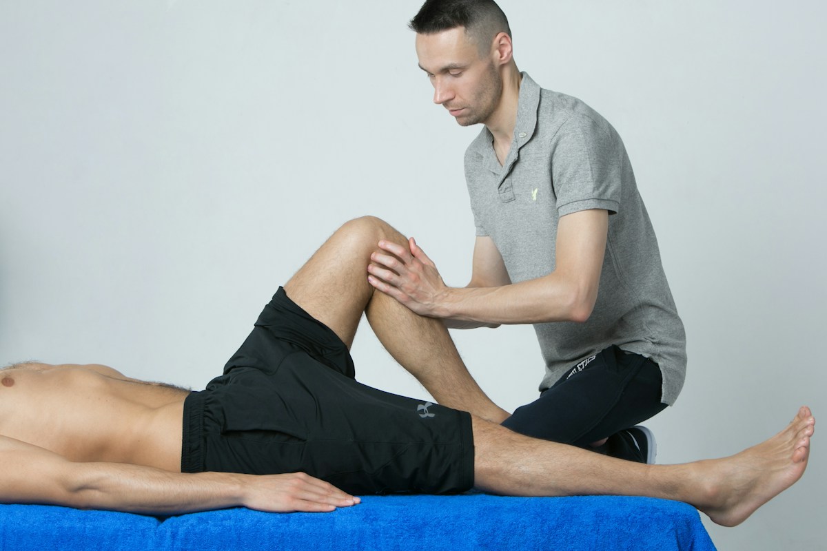
Tendinopathy represents one of the most pervasive, debilitating musculoskeletal disorders encountered in modern sports medicine, orthopedics, and clinical physical therapy, affecting elite athletic populations and sedentary individuals alike. Characterized clinically by localized, load-dependent tendon pain, impaired mechanical load-bearing capacity, morning stiffness, and profound functional disability, tendinopathy predominantly afflicts high-demand energy storage and transmission structures, including the patellar tendon, Achilles tendon, common extensor tendon of the lateral elbow, and the rotator cuff supraspinatus tendon. For decades, clinical management was fundamentally misled by the erroneous assumption that tendinopathy represented an acute inflammatory condition (‘tendinitis’), prompting the widespread, indiscriminate use of oral non-steroidal anti-inflammatory drugs and intratendinous corticosteroid injections.
Modern histopathological, biochemical, and biomechanical investigations have decisively dismantled the classical inflammatory paradigm, revealing that chronic tendon pathology is characterized not by prostaglandin-driven inflammatory cell infiltration, but by a non-inflammatory, cell-mediated degenerative cascade termed tendinosis. Ultrastructurally, tendinopathy exhibits profound ground substance matrix expansion, hypercellular tenocyte proliferation, hypervascular neovascular ingrowth with accompanying sensory nerve sprouting, and extensive disruption of the highly organized, parallel type I collagen fibrillar architecture. Consequently, modern clinical rehabilitation has undergone a radical transformation toward targeted mechanotherapy – utilizing precise mechanical loading protocols to stimulate cellular mechanotransduction, induce matrix remodeling, and restore tensile tendon capacity.
This comprehensive clinical guide provides an exhaustive analysis of neuromuscular tendinopathy rehabilitation, examining the continuum model of tendon pathology, tenocyte mechanobiology, the neurophysiology of tendon-induced cortical inhibition, and evidence-based loading paradigms. We detail the clinical application of heavy isometric loading for immediate analgesia, analyze heavy slow resistance (HSR) and eccentric loading for structural collagen cross-linking, and evaluate emerging adjunctive therapies, including autologous platelet-rich plasma (PRP) injections, extracorporeal shockwave therapy (ESWT), and high-volume image-guided hydrodissection, establishing a definitive roadmap for clinicians, orthopedic specialists, and physical therapists.
The Tendinopathy Continuum Model: Reactive, Dysrepair, and Degenerative Stages
To understand the clinical presentation and therapeutic responsiveness of tendon disorders, modern orthopedic science relies upon the Tendinopathy Continuum Model formulated by Cook and Purdam. This validated conceptual framework categorizes tendon pathology into three distinct, dynamically overlapping stages: reactive tendinopathy, tendon dysrepair (failed healing), and degenerative tendinopathy. Recognizing where an individual patient’s tendon resides along this pathological continuum is critical for determining appropriate mechanical loading parameters and avoiding interventions that exacerbate structural breakdown.
Reactive tendinopathy represents an acute, non-inflammatory cellular proliferative response to acute tensile or compressive overload, characteristically occurring when an unconditioned tendon experiences an abrupt surge in loading volume, frequency, or intensity. At the cellular level, tenocytes swell, becoming rounded and chondrocytic in appearance, and massively upregulate the synthesis of large, hydrophilic proteoglycans, predominantly aggrecan and versican. The intense negative electrical charge of these sulfated glycosaminoglycans draws abundant free water into the extracellular matrix, producing localized tendon thickening and tissue swelling that serves as an acute, short-term protective adaptation to disperse mechanical stress across a larger cross-sectional area.
If mechanical overload persists without adequate recovery intervals, the tendon transitions into the second stage: tendon dysrepair. During dysrepair, the rate of matrix breakdown overtakes endogenous cellular synthesis. Chondrocytic tenocytes synthesize abnormal, mechanically inferior type III collagen, which forms disorganized, irregular bundles that disrupt the parallel alignment of native type I collagen fibrils. Concurrently, localized microvascular ingrowth occurs, accompanied by perivascular sensory nerve fiber sprouting expressing substance P and calcitonin gene-related peptide (CGRP), which heighten localized nociceptive signaling. Importantly, tendon dysrepair remains partially reversible if mechanical load is promptly modified and appropriate mechanotherapy is introduced.
Degenerative tendinopathy represents the chronic, irreversible end-stage of tendon pathology, characterized by extensive, widespread tenocyte apoptosis, localized acellular necrotic areas, glassy hyaline degeneration, and complete structural disorganization of the collagen fibrillar matrix. Neovascularization and hyperinnervation are pronounced, while the remaining viable tendon matrix is crowded by disorganized type III collagen and fragmented elastin. Degenerative areas possess negligible tensile load-bearing capacity; under high-velocity stretch-shortening cycle loading, the concentrated mechanical strain placed upon the remaining thin rim of viable tendon tissue dramatically elevates the risk of acute, catastrophic tendon rupture.
Tenocyte Mechanobiology and Collagen Synthesis Kinetics
Tenocytes, the specialized elongated fibroblast-like cells of mesenchymal origin residing in longitudinal rows between parallel collagen bundles, function as the primary architects and mechanical sensors of tendon homeostasis. Quiescent tenocytes continuously monitor their microenvironment, regulating extracellular matrix turnover through the balanced expression of matrix metalloproteinases (MMPs) and tissue inhibitors of metalloproteinases (TIMPs). Mechanical loading serves as the obligate physiological stimulus that activates tenocytes to synthesize structural proteins.
Tenocyte mechanosensing is mediated primarily through cell surface integrin receptors (predominantly alpha-1/beta-1 and alpha-2/beta-1 integrins) that physically bridge extracellular collagen fibrils to the intracellular actin cytoskeleton, alongside stretch-activated ion channels (Piezo1, Piezo2, and TRPV4) and primary cilia projecting from the tenocyte membrane. When physical tensile strain stretches the tendon matrix, mechanical deformation opens stretch-activated calcium channels, inducing an immediate influx of extracellular calcium ions into the tenocyte cytoplasm. This cytosolic calcium transient activates calmodulin-dependent protein kinases and focal adhesion kinase (FAK), initiating intracellular signaling cascades.
The primary downstream transcriptional cascade driving tendon collagen synthesis is orchestrated by transforming growth factor-beta 1 (TGF-b1) and connective tissue growth factor (CTGF/CCN2). Active FAK and calcium signaling stimulate the phosphorylation and nuclear translocation of Smad2 and Smad3 transcription factors, inducing the transcription of genes encoding procollagen type I alpha-1 (COL1A1) and alpha-2 (COL1A2) chains. Inside the endoplasmic reticulum and Golgi apparatus of tenocytes, procollagen peptide chains undergo extensive post-translational modifications, including the hydroxylation of proline and lysine residues catalyzed by prolyl 4-hydroxylase and lysyl hydroxylase – enzymes that strictly require molecular oxygen, ferrous iron, and ascorbic acid (vitamin C) as indispensable cofactors.
Following cellular secretion into the extracellular space, specific procollagen N- and C-proteinases cleave the terminal globular registration peptides, yielding tropocollagen triple helices. Tropocollagen molecules spontaneously self-assemble into staggered microfibrils, fibrils, and fibers. The tensile strength and shear resistance of the mature tendon matrix are achieved through covalent inter- and intra-molecular cross-linking catalyzed by the extracellular enzyme lysyl oxidase (LOX). LOX oxidizes specific lysine and hydroxylysine residues into reactive aldehydes, forming divalent cross-links that mature into trivalent enzymatic cross-links (hydroxylysinonorleucine and pyridinoline), creating a robust, degradation-resistant fibrillar network capable of withstanding immense tensile forces.
The Neurophysiology of Tendon Pain and Cortical Inhibition
A profound breakthrough in modern sports neurology and physical therapy is the recognition that chronic tendinopathy is not merely a localized peripheral tissue injury, but a complex neurosensory and motor system disorder involving peripheral nociceptive sensitization, spinal cord hyper-reflexia, and profound cortical motor inhibition. Understanding this centralized neuromuscular re-organization explains why isolated passive treatments targeting the tendon matrix invariably fail to restore athletic performance and resolve functional deficits.
At the peripheral tissue level, tendinopathy pain is driven by localized biochemical and neurovascular abnormalities rather than classical inflammatory prostaglandins. In chronically degenerate tendon tissue, microvascular neovessels originating from the paratenon are accompanied by unmyelinated sensory nerve fibers that express high concentrations of nociceptive neurotransmitters, including substance P, glutamate, and calcitonin gene-related peptide (CGRP). These neuropeptides directly stimulate peripheral nociceptors and induce neurogenic vasodilation, generating a state of persistent peripheral nociceptive sensitization where even low, physiological mechanical loads provoke intense pain.
Ascending nociceptive input from the sensitized tendon induces secondary neuroplastic changes within the central nervous system. At the spinal cord level, dorsal horn sensory neurons exhibit central sensitization, lowering nociceptive thresholds and expanding receptive fields, manifesting clinically as primary and secondary hyperalgesia. More critically, transcranial magnetic stimulation (TMS) studies have revealed that chronic tendinopathy alters motor cortex neurophysiology. The primary motor cortex (M1) exhibits elevated intracortical inhibition and altered corticospinal excitability governing the muscle-tendon unit.
This motor cortex inhibition acts as a protective central nervous system ‘braking mechanism’ designed to prevent the patient from generating high, potentially destructive forces through the injured tendon. However, this profound cortical inhibition disrupts normal motor unit recruitment, induces muscle atrophy, and degrades inter-muscular coordination, creating an altered movement pattern that persists long after peripheral tendon pain has diminished. Consequently, effective tendinopathy rehabilitation must incorporate externally paced neuroplastic training modalities (such as auditory metronome pacing) to drive motor cortex neuroplasticity, reduce intracortical inhibition, and restore voluntary motor drive.
Heavy Isometric Loading: Neuroplastic Analgesia and Load Titration
The clinical implementation of heavy isometric loading represents one of the most significant breakthroughs in modern tendinopathy management, pioneered by Rio, Cook, and colleagues. In patients suffering from acute tendon irritability or severe load-related pain, performing dynamic exercises frequently exacerbates tendon symptoms secondary to high-velocity energy storage and release within the compromised tendon matrix. Isometric loading provides a safe, highly effective modality to apply high mechanical tension to tenocytes without exposing the tendon to cyclical stretch-shortening cycle strain.
The primary clinical utility of heavy isometric exercise lies in its remarkable, immediate analgesic potency. A landmark randomized crossover trial published in the British Journal of Sports Medicine demonstrated that performing 5 sets of 45-second heavy isometric contractions at 70 to 80 percent of maximal voluntary contraction (MVC), with 2-minute inter-set rest intervals, produced immediate, profound tendon pain relief lasting up to 45 to 60 minutes. Furthermore, concurrent transcranial magnetic stimulation revealed that this heavy isometric protocol immediately abolished cortical muscle inhibition, restoring maximal voluntary isometric force output.
The physiological mechanism underlying isometric-induced tendon analgesia involves the intense, sustained activation of large-diameter group Ib proprioceptive afferents originating from Golgi tendon organs and muscle spindles. High-threshold proprioceptive afferent barrage delivers powerful inhibitory presynaptic and postsynaptic signals to interneurons within the spinal cord dorsal horn, closing the pain gate under the Melzack-Wall gate control theory. Concurrently, heavy isometric holds stimulate the release of endogenous opioids and endocannabinoids within the periaqueductal gray (PAG) matter of the brainstem, activating descending noradrenergic and serotonergic pain-inhibitory pathways.
In clinical rehabilitation protocols, heavy isometric contractions (such as leg extensions for patellar tendinopathy or isometric heel raises for Achilles tendinopathy) serve as the essential foundational Phase 1 intervention. Clinicians prescribe 4 to 5 sets of 30 to 45-second holds, performed two to three times daily, utilizing isometric exercise as an ‘exercise-induced analgesic’ prior to sports training or physical therapy sessions, effectively controlling pain and enabling patients to engage in progressive rehabilitation without triggering reactive tendon flare-ups.
Heavy Slow Resistance and Eccentric Protocols: Collagen Cross-Linking
Once acute tendon irritability is stabilized using isometric loading, rehabilitation must transition toward progressive isotonic loading to stimulate tenocyte collagen synthesis, enhance tendon stiffness, and restore muscular capacity across the entire kinetic chain. Two primary isotonic loading paradigms have demonstrated exceptional, evidence-based efficacy in clinical trials: the Alfredson isolated eccentric protocol and the modern Heavy Slow Resistance (HSR) training paradigm developed by Kongsgaard and colleagues.
The Alfredson protocol, historically established for chronic midportion Achilles tendinopathy, focuses exclusively on isolated eccentric lengthening contractions, prescribing 3 sets of 15 repetitions performed twice daily with both straight-knee and bent-knee positions for 12 continuous weeks. Eccentric contractions subject the tendon to supramaximal mechanical strain, elongating muscle-tendon fascicles and applying high shear stress that physically disrupts pathological neovascular-nerve complexes. While highly effective, the isolated eccentric model suffers from poor long-term patient compliance due to the extraordinarily high time commitment (180 repetitions daily) and frequently induces marked muscle soreness.
The Heavy Slow Resistance (HSR) paradigm has largely superseded isolated eccentric protocols as the preferred clinical standard. HSR utilizes combined concentric and eccentric loading performed with heavy loads (beginning at 15-repetition maximum and progressing systematically to 6RM across 12 weeks), executed with a strict, slow tempo: 3 seconds concentric contraction, 0 seconds pause, and 3 seconds eccentric contraction (total 6 seconds per repetition). HSR is performed only three times per week, allowing essential 48-hour recovery intervals between training bouts.
In a landmark randomized clinical trial comparing HSR to the Alfredson eccentric protocol in patellar tendinopathy, HSR achieved equivalent long-term clinical pain reduction and VISA scores, but demonstrated superior patient compliance and higher patient satisfaction. More critically, high-resolution ultrasonography and tissue biopsies confirmed that HSR induced significant, measurable structural remodeling: tendon cross-sectional area decreased, abnormal neovascularization regressed, and tendon stiffness (Young’s modulus) increased substantially, driven by robust tenocyte procollagen type I synthesis and enzymatic collagen cross-linking stimulated by the slow, continuous tensile strain.
Energy Storage and Kinetic Chain Rehabilitation
A fundamental error in conventional physical therapy is discharging a patient after achieving painless isometric or slow resistance strength, without restoring the tendon’s primary biomechanical function: storing and releasing elastic energy during high-velocity locomotion. Tendons function as biological springs; during activities such as sprinting, jumping, hopping, and change of direction, the muscle contracts quasi-isometrically while the compliant tendon rapidly lengthens and recoils, storing kinetic energy during the eccentric braking phase and releasing it catapult-like during the concentric propulsion phase.
Restoring elastic energy storage capacity requires a dedicated Phase 3 rehabilitation protocol incorporating progressive stretch-shortening cycle (SSC) exercises. Because fast stretch-shortening cycle activities place immense mechanical strain on the tendon (e.g., patellar tendon loads reach 6 to 8 times body weight during jumping; Achilles tendon loads exceed 8 to 10 times body weight during sprinting), SSC exercises must be introduced systematically. Clinicians begin with low-amplitude, bilateral hopping drills, progressing to continuous skipping, bounding, box jumps, depth jumps, and sport-specific cutting maneuvers, ensuring that loading is performed every third day to permit 72 hours of collagen synthesis between sessions.
Furthermore, comprehensive tendinopathy rehabilitation mandates rigorous evaluation and conditioning of the entire kinetic chain. Tendons do not function in isolation; force transmission and load attenuation are shared across adjacent joint complexes and synergistic muscle groups. In Achilles tendinopathy, deficits in soleus muscle strength, quadriceps peak torque, and gluteus medius stabilization severely alter lower extremity landing biomechanics, shifting excessive decelerative forces onto the Achilles tendon.
Similarly, in patellar tendinopathy (jumper’s knee), restricted ankle dorsiflexion range of motion, gluteal neuromuscular inhibition, and poor eccentric quadriceps control force the knee into excessive valgus collapse and deep flexion during landing, dramatically amplifying patellar tendon tensile strain. Kinetic chain conditioning – including heavy soleus calf raises, Romanian deadlifts for posterior chain capacity, multi-planar hip abduction strengthening, and biomechanical landing retraining – distributes landing forces evenly across the entire musculoskeletal system, permanently offloading the vulnerable tendon.
Anatomical Variations: Insertional vs Midportion Tendon Pathomechanics
A fundamental diagnostic and therapeutic distinction in sports medicine is differentiating between midportion tendinopathy and insertional tendinopathy, as the underlying mechanical forces, histological features, and rehabilitation constraints diverge significantly between these two anatomical entities. While midportion tendinopathy is driven almost exclusively by excessive tensile loading and high-velocity stretch-shortening cycle strain, insertional tendinopathy is characterized by the complex interaction of both tensile forces and compressive mechanical shear against bony prominences.
Insertional Achilles tendinopathy occurs within the distal 2 centimeters of the tendon at its enthesis onto the posterior calcaneus. In this anatomical zone, the deep surface of the tendon is subjected to repetitive mechanical compression against the posterosuperior calcaneal tuberosity during ankle dorsiflexion. This compressive impingement is frequently exacerbated by anatomical variations, such as a prominent Haglund’s deformity (retrocalcaneal exostosis), and is accompanied by inflammatory involvement of the adjacent retrocalcaneal bursa (retrocalcaneal bursitis) and superficial subcutaneous bursa.
Under continuous compressive stress, enthesis tenocytes undergo chondroid metaplasia, synthesizing fibrocartilage and abundant aggrecan proteoglycans that mineralize into painful intratendinous calcifications and traction osteophytes. Clinically, recognizing insertional pathology dictates an absolute modification of rehabilitation protocols: loaded dorsiflexion past a neutral 0-degree angle must be strictly avoided during early and intermediate loading phases. Unlike midportion tendinopathy, where heel raises are performed off the edge of a step through full dorsiflexion, insertional Achilles exercises must be performed exclusively from the flat floor to eliminate destructive compressive impingement against the calcaneal spur.
Similarly, in proximal patellar tendinopathy (jumper’s knee), mechanical impingement occurs at the deep, posterior interface of the patellar tendon against the inferior pole of the patella during deep knee flexion (> 60 to 90 degrees). In the flexed knee position, tensile strain concentrates on the anterior tendon fibers while compressive shear deforms the posterior fibers against the patellar bone. Rehabilitation protocols must therefore utilize decline slant board squats restricted to 0 to 60 degrees of flexion during initial phases, avoiding deep knee bending until compressive remodeling tolerance has been restored.
Nutritional Mechanobiology: Gelatin, Vitamin C, and Collagen Synthesis Kinetics
While mechanical loading provides the primary mechanical stimulus for tenocyte mechanotransduction, nutritional availability dictates the biochemical rate of procollagen monomer synthesis and extracellular matrix cross-linking. Tendon collagen consists of a repeating triplet amino acid sequence – Glycine-X-Y, where X and Y are predominantly L-proline and L-hydroxyproline. Because glycine constitutes exactly one-third of all amino acids in the collagen triple helix, and proline/hydroxyproline account for another 20 to 25 percent, endogenous synthesis can be limited by the biological availability of these non-essential and semi-essential amino acids during periods of intense rehabilitative loading.
Groundbreaking research pioneered by Keith Baar and colleagues at the University of California, Davis, has established a targeted nutritional mechanobiology protocol designed to maximize tendon collagen synthesis. When athletes ingest 15 grams of pharmaceutical-grade gelatin or hydrolyzed collagen peptides accompanied by 50 milligrams of vitamin C (ascorbic acid) exactly 30 to 60 minutes prior to a brief (5 to 10-minute) mechanical loading session, circulating plasma concentrations of glycine, proline, hydroxyproline, and hydroxylysine peak precisely when mechanical strain activates tenocyte cellular machinery.
Ascorbic acid serves as an indispensable electron-donating cofactor for the endoplasmic reticulum enzymes prolyl 4-hydroxylase and lysyl hydroxylase. These enzymes catalyze the post-translational hydroxylation of proline and lysine residues on nascent procollagen polypeptide chains, an obligate biochemical step required to form the inter-chain hydrogen bonds that stabilize the collagen triple helix. Deficiency of vitamin C completely halts procollagen folding and triggers intracellular protein degradation, preventing extracellular secretion.
Clinical biomarker trials evaluating this targeted nutritional intervention demonstrated that ingesting gelatin and vitamin C prior to intermittent jump rope or isometric loading doubled the circulating concentrations of procollagen type I amino-terminal propeptide (PINP) – the primary serum biomarker of systemic bone and tendon collagen formation. Incorporating this pre-exercise nutritional strategy into chronic tendinopathy rehabilitation programs accelerates structural tissue repair, enhances tendon tensile modulus, and shortens the clinical timeline required for safe return to competitive sport.
Biomechanical Load Monitoring and the 24-Hour Pain Response
A critical clinical challenge in managing tendinopathy is navigating the delayed biological response of tendon tissue to mechanical loading. Tendons lack the dense capillary networks and rapid sensory feedback mechanisms of skeletal muscle; consequently, a tendon may feel completely pain-free or mildly stiff during a strenuous athletic workout, only to develop severe, disabling pain and reactive swelling 12 to 24 hours later. This delayed response frequently lures athletes and clinicians into a vicious cycle of overtraining followed by acute flare-ups.
To eliminate this diagnostic uncertainty, modern sports medicine utilizes the validated ’24-Hour Pain Response’ monitoring protocol. Under this monitoring algorithm, the patient identifies a standardized, highly reproducible, sport-specific tendon loading test – such as a single-leg decline squat on a 25-degree slant board for patellar tendinopathy, a single-leg heel raise or hop for Achilles tendinopathy, or a gripping test for lateral elbow tendinopathy. This test is performed once daily at the exact same time every morning (typically within 30 minutes of waking).
The patient rates their pain on a numeric rating scale from 0 to 10 during the morning loading test. Clinical decision-making is guided by strict thresholds: pain levels up to 3 to 4 out of 10 during the morning test are acceptable and indicate that the tendon is safely adapting to the previous day’s mechanical loading, provided that the pain returns to baseline within 24 hours. However, if morning pain jumps to 5 or higher, or if morning stiffness duration increases noticeably compared to baseline, it indicates that the previous day’s load exceeded the tendon’s current mechanical capacity, mandating an immediate reduction in training intensity and volume back to the previous tolerated loading stage.
Diagnostic Ultrasonography and Ultrasound Tissue Characterization (UTC)
Accurate diagnostic imaging and longitudinal structural monitoring are vital components of modern tendinopathy management, enabling sports medicine physicians to assess pathological severity, rule out partial tears, and track tissue recovery. Conventional greyscale ultrasound (B-mode) remains the first-line imaging modality, demonstrating pathognomonic features of tendinosis: localized fusiform tendon thickening, hypoechoic areas representing focal collagen disorganization and ground substance expansion, loss of the normal fibrillar echotexture, and cortical irregularity at the bony insertion.
Power Doppler ultrasonography provides qualitative evaluation of pathological neovascularization. In asymptomatic, healthy tendons, intrinsic blood flow is minimal and undetectable on standard Doppler imaging. In contrast, symptomatic tendinopathic tendons exhibit marked localized hypervascularity, where low-velocity blood flow through pathological neovessels is visualized infiltrating the deep tendon substance. Importantly, clinicians must recognize that the degree of Doppler neovascularization does not correlate directly with clinical pain severity, and persistent neovascularity can remain visible on ultrasound even after complete symptomatic resolution.
Ultrasound Tissue Characterization (UTC) represents an advanced, objective imaging technology developed specifically to overcome the subjective observer variability of conventional B-mode ultrasound. UTC utilizes a motorized tracking transducer that captures standardized transverse ultrasound images every 0.2 millimeters along the longitudinal axis of the tendon. Proprietary tomographic algorithms analyze the stability of acoustic reflection patterns across contiguous images, categorizing tendon tissue into four distinct, color-coded ‘Echo Types’.
Echo Type I (green) represents intact, highly organized parallel collagen bundles; Echo Type II (blue) denotes discontinuous, wavy, or swollen collagen fibrils; Echo Type III (red) corresponds to disorganized fibrillar matrix with abundant proteoglycans and free water; and Echo Type IV (black) represents amorphous, acellular necrotic matrix and fluid. By providing quantitative percentages of each Echo Type across the entire tendon volume, UTC allows clinicians to objectively evaluate matrix response to training loads, verify that heavy resistance protocols are increasing Type I green collagen without destabilizing the matrix into Type III or IV, and make evidence-based return-to-play decisions.
Biologic Adjuncts: Platelet-Rich Plasma, Shockwave, and Hydrodissection
In patients with chronic, recalcitrant tendinopathy who fail to achieve functional recovery despite 3 to 6 months of structured, progressive mechanotherapy, biologic and minimally invasive procedural adjuncts can be integrated into the treatment paradigm. However, clinicians must critically evaluate the clinical trial evidence and understand the precise biological mechanisms of these modalities to avoid ineffective or potentially destructive interventions.
Extracorporeal Shockwave Therapy (ESWT), delivering focused or radial acoustic shockwaves into the degenerated tendon substance, is one of the most thoroughly validated non-invasive adjuncts. High-energy acoustic waves induce localized mechanical cavitation, stimulating microvascular perfusion and upregulating tenocyte expression of proliferating cell nuclear antigen (PCNA), TGF-b1, and VEGF. Furthermore, ESWT selectively depletes substance P and calcitonin gene-related peptide from sensory nerve endings within the tendon, delivering clinically meaningful, long-term pain reduction. Randomized clinical trials have confirmed that combining ESWT with heavy slow resistance training achieves significantly higher success rates than either modality alone.
Autologous Platelet-Rich Plasma (PRP) therapy involves centrifuging whole blood to concentrate platelets 4 to 8 times above baseline, subsequently injecting the platelet concentrate directly into the degenerated tendon under continuous real-time ultrasound guidance. Alpha granules within platelets release a dense cocktail of anabolic growth factors – including platelet-derived growth factor (PDGF), vascular endothelial growth factor (VEGF), transforming growth factor-beta 1, and basic fibroblast growth factor (bFGF) – that stimulate tenocyte proliferation and extracellular matrix synthesis. While clinical trials show mixed results in unselected populations, leukocyte-poor PRP (LP-PRP) has demonstrated clear clinical superiority over corticosteroids in chronic patellar and lateral epicondylar tendinopathy.
High-Volume Image-Guided Injection (HVIGI), or hydrodissection, represents an exceptional, minimally invasive interventional modality for chronic Achilles and patellar tendinopathy characterized by prominent neovascularization. Under continuous ultrasound guidance, a physician injects a high volume (30 to 50 mL) of sterile saline containing small concentrations of local anesthetic into the pathological interface between the anterior aspect of the Achilles tendon and Kager’s fat pad. The high hydrostatic pressure mechanically shears and obliterates the fragile neovascular-nerve complexes responsible for chronic pain, delivering immediate, dramatic symptom relief and enabling patients to resume heavy loading protocols.
To assist sports medicine physicians, orthopedic physical therapists, and athletic training staff in navigating the complex continuum of tendon rehabilitation, the following comparative clinical matrix details the primary mechanotherapeutic loading stages and procedural adjuncts. Each intervention is delineated by its underlying mechanical stimulus, biological target, execution tempo, and guideline-endorsed clinical timing.
Adhering to this structured, progressive loading continuum ensures that mechanical stress is precisely matched to current tendon capacity, preventing catastrophic tendon rupture while driving long-term collagen remodeling and restoring athletic performance.
| Rehabilitation Stage / Modality | Mechanical Stimulus / Loading Protocol | Biological & Neurophysiological Impact | Key Clinical Evidence | Clinical Timing & Criteria |
|---|---|---|---|---|
| Phase 1: Heavy Isometric Loading | 4-5 sets x 30-45s holds @ 70-80% MVC; 2 min rest; 2-3 times daily | Immediate pain analgesia (45 min); abolishes cortical motor inhibition; no tendon strain | Rio et al. (BJSM 2015), van Ark et al. | Initial phase; acute reactive flares; high morning pain (> 4/10) |
| Phase 2: Heavy Slow Resistance (HSR) | 3-4 sets x 6-10RM @ 3s concentric / 3s eccentric tempo; 3 days/week | Stimulates tenocyte procollagen synthesis; LOX cross-linking; increases tendon stiffness | Kongsgaard et al. (Scand J Med Sci Sports) | Initiated once isometric pain is stable; foundational structural remodeling phase |
| Phase 3: Stretch-Shortening Cycle (SSC) | Progressive plyometrics (hopping, bounding, depth jumps); every 3rd day | Restores spring-like elastic energy storage & release; high peak rate of force development | Silbernagel et al., Malliaras et al. | Requires limb strength symmetry >= 85% and pain-free HSR loading |
| Phase 4: Sport-Specific Return to Play | Full sprinting, decelerations, sport drills; 24-hour morning pain monitoring | Full functional integration across kinetic chain; dynamic neuromuscular motor control | Cook & Purdam (BJSM Consensus) | Full sport participation; maintained maintenance isometric/HSR training |
| Extracorporeal Shockwave Therapy (ESWT) | Focused acoustic shockwaves (2000-3000 pulses @ 1.5-2.5 bar, 4-8 Hz; 3-5 sessions) | Cavitation-induced tenocyte activation; neuropeptide (Substance P) depletion | Rompe et al., Vetrano et al. (Am J Sports Med) | Adjunct for chronic calcific or non-responsive tendinopathy > 3 months |
| High-Volume Image-Guided Hydrodissection | Ultrasound-guided injection of 30-50 mL saline + local anesthetic into Kager’s space | Mechanical shearing and destruction of pain-producing neovascular-nerve bundles | Chan et al., Wheeler et al. | Refractory Achilles/patellar tendinopathy with confirmed high neovascularization |
The structured rehabilitative progression presented above highlights the vital importance of staged mechanotherapy in managing tendon disorders. Rather than resting or relying on passive modalities, prescribing precise, progressive mechanical loads restores tenocyte metabolism, reconstructs collagen alignment, and allows athletes to safely return to full competitive play.
Frequently Asked Questions About Tendinopathy Rehabilitation
Why is tendinopathy considered a degenerative condition rather than an inflammatory ‘tendinitis’?
Extensive histological and biochemical studies confirm that chronic tendinopathy lacks classical inflammatory cells (neutrophils and lymphocytes) and elevated prostaglandins. Instead, the tissue exhibits non-inflammatory degenerative changes, including disorganized type III collagen, increased proteoglycans and water, tenocyte hypercellularity, and abnormal neovascularization with pain-sensing nerve sprouting. Consequently, anti-inflammatory treatments like corticosteroids do not cure the condition and can worsen tendon degeneration.
How does heavy isometric loading provide immediate tendon pain relief?
Heavy isometric loading (5 sets of 45-second holds at 70-80% maximal voluntary contraction) delivers an intense proprioceptive afferent signal through Golgi tendon organs and muscle spindles. This stimulates inhibitory interneurons in the spinal cord dorsal horn to close the pain gate, while simultaneously activating descending pain-inhibitory pathways in the brainstem and abolishing cortical motor inhibition, providing pain relief lasting 45 to 60 minutes.
What is Heavy Slow Resistance (HSR) training and why is it superior to fast lifting?
Heavy Slow Resistance involves performing combined concentric and eccentric exercises with heavy loads (6-10RM) at a strict, slow tempo (3 seconds up, 3 seconds down) three times per week. The slow tempo eliminates momentum, subjecting tenocytes to sustained tensile strain that stimulates procollagen type I synthesis and enzymatic collagen cross-linking, resulting in increased tendon stiffness and long-term structural remodeling.
How does the 24-hour pain response guide tendon rehabilitation?
Because tendons respond to overload with a delayed pain response, athletes monitor localized pain during a standardized test (such as a single-leg decline squat) every morning upon waking. Pain up to 3-4 out of 10 that settles within 24 hours indicates safe loading. However, if morning pain reaches 5 or higher, or morning stiffness increases significantly, it signals that the previous day’s load exceeded tendon capacity, requiring training modification.
What is the role of the stretch-shortening cycle (SSC) in tendon function?
Tendons act as biological springs, storing and releasing elastic energy during high-velocity stretch-shortening cycle activities like sprinting and jumping. Rehabilitation must progress from slow strength training to fast plyometric and hopping exercises to restore the tendon’s elastic recoil capacity, which cannot be achieved through slow resistance training alone.
Why are corticosteroid injections harmful for chronic tendinopathy?
While corticosteroid injections provide short-term pain relief through non-specific membrane stabilization, they are directly cytotoxic to tenocytes. Corticosteroids suppress collagen synthesis, induce tenocyte necrosis, and significantly reduce tendon mechanical strength, resulting in extraordinarily high recurrence rates and a substantially increased risk of tendon rupture.
How does high-volume image-guided hydrodissection relieve Achilles tendon pain?
Hydrodissection involves injecting 30 to 50 mL of sterile saline under ultrasound guidance into the interface between the Achilles tendon and Kager’s fat pad. The high fluid volume mechanically shears and destroys the pathological neovascular network and accompanying sensory nerve fibers that sprout into the degenerate tendon, delivering rapid pain relief.
Why must the kinetic chain be addressed in tendinopathy rehabilitation?
Tendons operate as part of an integrated musculoskeletal chain. Weakness or neuromuscular deficits in adjacent muscles (such as the soleus in Achilles tendinopathy, or the quadriceps and gluteals in patellar tendinopathy) impair shock absorption during landing, shifting excessive decelerative forces directly onto the vulnerable tendon. Conditioning the entire kinetic chain distributes forces evenly and prevents tendon overload.
Clinical Perspectives and Future Directions in Tendon Sports Medicine
The clinical management of tendinopathy has advanced from empirical rest and passive modalities into a precise biological science anchored in tenocyte mechanotransduction, neuromuscular neuroplasticity, and progressive loading. By understanding the multi-stage continuum of tendon pathology, sports clinicians and physical therapists can prescribe specific loading profiles that match the biological capacity of the tissue.
Systematically progressing patients through heavy isometric loading for analgesia, heavy slow resistance training for collagen matrix cross-linking, and fast stretch-shortening cycle exercises for elastic energy storage provides a comprehensive framework that restores full functional capacity. Coupled with kinetic chain conditioning and meticulous 24-hour morning pain monitoring, athletes can achieve durable recovery and prevent future tendon breakdown.
For accredited institutional guidelines and evidence-based sports medicine consensus, clinicians are encouraged to consult clinical recommendations published by the British Journal of Sports Medicine (BJSM), the Journal of Orthopaedic & Sports Physical Therapy (JOSPT), and research literature from the American Orthopaedic Society for Sports Medicine. Landmark clinical trial data are continuously cataloged on PubMed National Library of Medicine, alongside musculoskeletal rehabilitation perspectives from the World Health Organization.
