


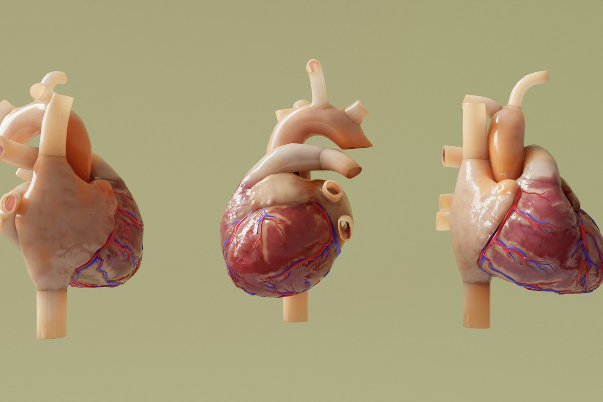
The vascular endothelium, a contiguous monocellular monolayer lining the entire human circulatory tree, represents far more than an inert anatomical barrier separating circulating blood from underlying vascular smooth muscle; it functions as a highly sophisticated, autocrine, paracrine, and endocrine organ that continuously modulates vascular tone, platelet aggregation, leukocyte adhesion, and vascular smooth muscle proliferation. At the center of endothelial homeostatic regulation lies nitric oxide (NO), a lipophilic, gaseous signaling molecule whose discovery revolutionized vascular biology and cardiovascular medicine.
Endothelial dysfunction, characterized by the progressive loss of bioavailable nitric oxide, represents the earliest detectable subclinical pathological precursor in the pathogenesis of atherosclerosis, essential hypertension, coronary artery disease, and diabetic microvascular angiopathy. Loss of NO bioavailability is driven by a convergence of biochemical perturbations: eNOS uncoupling, depletion of the essential cofactor tetrahydrobiopterin (BH4), accumulation of the endogenous competitive inhibitor asymmetric dimethylarginine (ADMA), and excessive generation of superoxide free radicals that scavenge NO to form cytotoxic peroxynitrite.
This comprehensive clinical treatise delivers an exhaustive examination of vascular endothelial physiology and nitric oxide biology. We analyze the dual pathways of systemic NO generation – the classical L-arginine-eNOS cascade and the alternative nitrate-nitrite-nitric oxide enterosalivary pathway, dissect the biophysical mechanotransduction of fluid shear stress and flow-mediated dilation (FMD), evaluate clinical evidence regarding dietary inorganic nitrate supplementation and oral microbiome symbiosis, and review therapeutic strategies designed to restore endothelial glycocalyx integrity and optimize lifelong cardiovascular longevity.
The Vascular Endothelium as a Dynamic Endocrine Organ: Homeostatic Roles
Spanning an estimated surface area of over 4,000 square meters in the adult human body, the vascular endothelium represents one of the largest and most metabolically active endocrine interfaces in mammalian physiology. Composed of approximately one trillion endothelial cells lining arteries, arterioles, capillaries, venules, and veins, the healthy endothelium maintains cardiovascular homeostasis through continuous paracrine signaling.
The phenotypic state of the healthy endothelium is fundamentally anti-thrombotic, anti-inflammatory, and vasodilatory. Endothelial cells continuously synthesize and release an array of bioactive autacoids, including nitric oxide, prostacyclin (PGI2), and endothelium-derived hyperpolarizing factor (EDHF / epoxyeicosatrienoic acids), counterbalanced by vasoconstrictor substances such as endothelin-1 (ET-1), thromboxane A2 (TxA2), and angiotensin II.
In physiological health, the vasodilatory and protective signaling cascades dominate, maintaining low systemic vascular resistance, inhibiting platelet activation and aggregation, preventing leukocyte rolling and transmigration, and suppressing vascular smooth muscle cell phenotypic switching and neointimal hyperplasia.
However, when exposed to chronic hemodynamic perturbations, hyperlipidemia, cigarette smoke toxins, hyperglycemia, or systemic inflammatory cytokines, the endothelium undergoes phenotypic activation and progressive decay termed endothelial dysfunction.
Endothelial dysfunction is defined pathologically as the loss of endothelium-dependent vasodilation, primarily secondary to diminished bioavailable nitric oxide, shifting the vascular wall into a pro-thrombotic, pro-inflammatory, and vasoconstrictive state that initiates atherosclerotic plaque formation.
eNOS Enzymology and Caveolar Localization: The L-Arginine Oxidation Pathway
Under physiological conditions, the primary endogenous source of vascular nitric oxide is endothelial nitric oxide synthase (eNOS / NOS3), a constitutive, homodimeric, calcium/calmodulin-dependent hemoprotein enzyme tightly associated with the plasma membrane.
Functionally active eNOS exists as a homodimer; each monomer consists of a C-terminal reductase domain (binding NADPH, flavin adenine dinucleotide [FAD], and flavin mononucleotide [FMN]) and an N-terminal oxygenase domain (binding iron protoporphyrin IX heme, the essential pterin cofactor (6R)-5,6,7,8-tetrahydrobiopterin [BH4], and the substrate L-arginine).
In resting endothelial cells, eNOS is localized to specialized, cholesterol- and sphingolipid-rich invaginations of the plasma membrane termed caveolae. Within caveolae, eNOS is maintained in an enzymatically inactive, inhibited state through tonic protein-protein binding with caveolin-1 (specifically its scaffolding domain, residues 82-101).
Upon endothelial stimulation by receptor agonists (acetylcholine, bradykinin, histamine) or mechanical fluid shear stress, intracellular calcium (Ca2+) surges, binding to calmodulin. The calcium-calmodulin complex binds with high affinity to the linker region of eNOS, competitively displacing caveolin-1 and relieving basal enzymatic inhibition.
Once activated, eNOS transfers electrons from NADPH through FAD and FMN in the reductase domain to the heme iron of the oxygenase domain. This electron flux catalyzes the five-electron oxidation of the guanidino nitrogen of L-arginine, proceeding through the intermediate N-omega-hydroxy-L-arginine (NOHLA) to produce stoichiometric quantities of nitric oxide and L-citrulline.
eNOS Uncoupling: BH4 Depletion and Superoxide Radical Production
Under pathological conditions characterized by elevated oxidative stress, endothelial nitric oxide synthase undergoes a catastrophic enzymatic transition known as ‘eNOS uncoupling’. Instead of synthesizing protective nitric oxide, uncoupled eNOS transforms into a dysfunctional generator of cytotoxic reactive oxygen species.
The central biochemical determinant of eNOS coupling status is the availability of its essential cofactor, tetrahydrobiopterin (BH4). BH4 acts not merely as a structural stabilizer of the active eNOS homodimer, but as an essential electron donor that participates directly in oxygen activation.
When endothelial cells are exposed to oxidative stressors (such as oxidized low-density lipoprotein, advanced glycation end-products, or angiotensin II), vascular NADPH oxidases (NOX2 and NOX4) are activated, generating high concentrations of superoxide anions (O2-). Superoxide rapidly reacts with endogenous nitric oxide to form peroxynitrite (ONOO-), an extraordinarily potent oxidant.
Peroxynitrite diffuses to eNOS and oxidizes the redox-sensitive zinc-thiolate cluster in the dimer interface, while simultaneously oxidizing tetrahydrobiopterin (BH4) into enzymatically inactive dihydrobiopterin (BH2) and biopterin. Because BH2 binds to the eNOS oxygenase domain with equal affinity to BH4 but is incapable of donating electrons, BH2 competitively displaces BH4.
In the absence of functional BH4, the flow of electrons through eNOS becomes ‘uncoupled’ from L-arginine oxidation. Electrons flowing from NADPH to the catalytic heme are transferred directly to molecular oxygen, reducing O2 to superoxide (O2-) rather than synthesizing nitric oxide. Consequently, uncoupled eNOS exacerbates the oxidative milieu, establishing a self-reinforcing vicious cycle of endothelial destruction.
The Alternative Nitrate-Nitrite-Nitric Oxide Enterosalivary Pathway
For over a century, the classical L-arginine-eNOS pathway was presumed to be the exclusive source of bioavailable nitric oxide in the human body. However, landmark translational discoveries in human physiology identified an alternative, non-enzymatic physiological pathway: the nitrate-nitrite-nitric oxide pathway.
Unlike the oxygen-dependent L-arginine pathway, which fails under conditions of hypoxia, acidosis, and endothelial damage, the nitrate-nitrite-NO pathway is oxygen-independent and is actively accelerated by low oxygen tensions and acidic pH, functioning as an essential backup system for vascular perfusion.
This pathway relies on the sequential biological reduction of inorganic nitrate (NO3-), obtained primarily from dietary plant sources, to inorganic nitrite (NO2-), and subsequently to bioavailable nitric oxide (NO).
Following ingestion, dietary inorganic nitrate is rapidly absorbed in the upper small intestine, entering systemic circulation. While approximately 70 percent of ingested nitrate is eventually excreted by the kidneys, the remaining 25 percent is actively extracted from the bloodstream by the salivary glands via active sodium-dependent sialin nitrate/iodide transporters (SLC17A5), concentrating nitrate in human saliva up to 10 to 20-fold compared to blood plasma.
This concentrated nitrate is continuously secreted into the oral cavity, initiating the obligate enterosalivary circulation that couples human cardiovascular health with the oral microbiome.
Oral Microbiome Symbiosis: Bacterial Nitrate Reductases in the Oral Cavity
Mammalian cells completely lack functional nitrate reductase enzymes capable of reducing inorganic nitrate (NO3-) into bioactive nitrite (NO2-). Consequently, the human body is entirely dependent on a symbiotic relationship with facultative and obligate anaerobic bacteria inhabiting the dorsal surface of the tongue and interdental crevices.
Within the deep crypts of the posterior tongue, low oxygen microenvironments harbor dense microbial communities, predominantly belonging to the genera Veillonella (Veillonella atypica, Veillonella dispar), Actinomyces (Actinomyces odontolyticus), Rothia (Rothia mucilaginosa), and Prevotella.
These oral commensal bacteria express functional membrane-bound nitrate reductase (Nar) enzyme complexes. The Nar complex utilizes dietary nitrate as an alternative terminal electron acceptor during anaerobic cellular respiration, enzymatically reducing nitrate (NO3-) to nitrite (NO2-) and expelling nitrite into the salivary flow.
Swallowed saliva delivers high concentrations of nitrite into the gastric lumen. In the highly acidic environment of the stomach (gastric juice pH 1.5 to 2.0), nitrite is immediately protonated to form nitrous acid (HNO2), which decomposes into nitric oxide, nitrogen dioxide, and reactive nitrogen intermediates that disinfect swallowed pathogens, stimulate gastric mucosal blood flow, and enhance protective gastric mucus secretion.
Residual unreduced nitrite passes into the duodenum, where it is absorbed into the bloodstream, establishing elevated circulating plasma nitrite reserves (typically 100 to 500 nmol/L) that circulate throughout the arterial tree as stable NO precursors.
Flow-Mediated Dilation: Biophysical Principles and High-Resolution Ultrasound
Flow-Mediated Dilation (FMD) represents the clinical gold standard, non-invasive diagnostic modality for evaluating endothelial nitric oxide bioavailability in human subjects, established by Celermajer and colleagues.
The biophysical principle of FMD relies on the physiological response of large conduit arteries (typically the brachial artery of the upper arm) to an acute increase in fluid shear stress following a period of transient downstream tissue ischemia.
Under standard clinical guidelines established by the International Brachial Artery Reactivity Task Force, a high-resolution vascular ultrasound probe (10 to 15 MHz) is positioned over the brachial artery 5 to 10 cm above the antecubital fossa. Baseline resting arterial lumen diameter is measured using automated edge-detection wall tracking software.
A pneumatic blood pressure occlusion cuff placed on the forearm is inflated to suprasystolic pressure (typically 200 to 250 mmHg, or 50 mmHg above resting systolic pressure) for exactly five minutes, inducing complete distal forearm ischemia. Upon rapid cuff deflation, downstream resistance arterioles undergo maximal metabolic vasodilation, provoking a massive surge in blood flow velocity through the conduit brachial artery termed ‘reactive hyperemia’.
The rapid flow of blood exerts elevated frictional drag (shear stress) against the luminal endothelial surface, stimulating immediate eNOS activation and nitric oxide release. NO diffuses into vascular smooth muscle, provoking arterial vasodilation that peaks 60 to 90 seconds post-deflation. FMD is expressed as the percentage change in peak diameter relative to baseline diameter: FMD (%) = [(Peak Diameter – Baseline Diameter) / Baseline Diameter] * 100. In healthy young adults, normal FMD values range between 7 and 12 percent; values < 5 to 6 percent indicate severe endothelial dysfunction and independently predict future cardiovascular events.
Hemodynamic Fluid Shear Stress: Mechanosensing and Cytoskeletal Transduction
Fluid shear stress – the tangential frictional force exerted by the laminar flow of viscous blood across the apical surface of endothelial cells, quantified in dynes per square centimeter (dyn/cm^2) – represents the primary physical stimulus directing endothelial gene expression, morphology, and nitric oxide production.
Endothelial cells detect mechanical shear stress through a sophisticated mechanosensory complex situated at endothelial cell-cell adherens junctions, composed of Platelet Endothelial Cell Adhesion Molecule-1 (PECAM-1 / CD31), Vascular Endothelial Cadherin (VE-cadherin), and Vascular Endothelial Growth Factor Receptor 2 (VEGFR2).
When laminar shear stress acts on the luminal cell surface, PECAM-1 acts as the primary mechanical sensor, undergoing conformational deformation that transmits tensile force to VE-cadherin. VE-cadherin functions as an adapter that clusters and transactivates VEGFR2 in a ligand-independent manner.
Phosphorylated VEGFR2 triggers downstream signaling through phosphatidylinositol 3-kinase (PI3K) and the serine/threonine protein kinase Akt (Protein Kinase B). Activated Akt directly phosphorylates eNOS at its critical activation site, serine residue 1177 (Ser1177). Phosphorylation of Ser1177 alters the C-terminal tail of eNOS, enhancing electron transfer flux from the reductase to the oxygenase domain and increasing baseline NO production by two- to four-fold, independent of intracellular calcium fluctuations.
Conversely, in regions of arterial branching, bifurcations, and curvatures, disturbed, turbulent, or low oscillatory shear stress (< 4 dyn/cm^2) fails to activate Akt, triggering instead pro-inflammatory NF-kB activation, VCAM-1 expression, and eNOS downregulation, explaining why atherosclerotic plaques develop selectively at arterial bends and bifurcations.
Vascular Smooth Muscle Relaxation: sGC, cGMP, and PKG Signaling Cascades
Once synthesized and released by endothelial cells, nitric oxide exerts its primary vasodilatory action by rapidly diffusing across the internal elastic lamina into adjacent vascular smooth muscle cells (VSMCs).
Because nitric oxide is a neutral, uncharged lipophilic molecule with a biological half-life of merely 1 to 5 milliseconds in blood, it crosses cell membranes via passive diffusion. Within vascular smooth muscle, NO binds with picomolar affinity to the prosthetic ferrous (Fe2+) heme moiety of soluble guanylyl cyclase (sGC), its primary intracellular receptor enzyme.
Binding of NO to sGC induces a conformational shift that breaks the axial iron-histidine bond, activating the catalytic domain of sGC by several hundred-fold. Activated sGC catalyzes the conversion of guanosine triphosphate (GTP) into the intracellular secondary messenger cyclic guanosine monophosphate (cGMP).
Elevated cGMP binds to and activates cyclic GMP-dependent protein kinase I (PKG-I / cGK-I). Activated PKG orchestrates vascular smooth muscle relaxation through multiple simultaneous molecular actions: phosphorylating phospholamban (PLN) to stimulate sarcoplasmic/endoplasmic reticulum calcium ATPase (SERCA2a), pumping cytosolic calcium back into internal sarcoplasmic stores; inhibiting L-type voltage-gated calcium channels (Cav1.2), halting extracellular calcium influx; and opening large-conductance calcium-activated potassium channels (BKCa), hyperpolarizing the plasma membrane.
Most crucially, PKG phosphorylates and activates myosin light chain phosphatase (MLCP). MLCP dephosphorylates the 20-kDa regulatory light chain of myosin (MLC20), dissociating actin-myosin cross-bridges and inducing immediate, profound vascular smooth muscle relaxation and luminal vasodilation.
Asymmetric Dimethylarginine: The Endogenous eNOS Competitive Antagonist
A major pathophysiological mechanism driving diminished nitric oxide bioavailability in cardiovascular and metabolic disease is the biological accumulation of asymmetric dimethylarginine (ADMA), an endogenous competitive inhibitor of eNOS.
ADMA is generated during the normal physiological turnover of intracellular proteins. Nuclear and cytoplasmic proteins undergo post-translational methylation of arginine residues by the enzyme family protein arginine methyltransferases (PRMTs, specifically Type I PRMTs). Upon lysosomal and proteasomal degradation of these methylated proteins, free ADMA and its stereoisomer symmetric dimethylarginine (SDMA) are released into the cytoplasm and bloodstream.
Because ADMA shares high structural homology with L-arginine, it competes with L-arginine for transport across cellular membranes via cationic amino acid transporters (CAT-1 / y+ system). Once inside endothelial cells, ADMA directly binds to the substrate-binding pocket in the oxygenase domain of eNOS, competitively displacing L-arginine and completely blocking nitric oxide synthesis.
Under normal physiological conditions, over 80 percent of generated ADMA is enzymatically metabolized and cleared by the intracellular enzyme dimethylarginine dimethylaminohydrolase (DDAH, isoforms DDAH-1 and DDAH-2), which hydrolyzes ADMA into L-citrulline and dimethylamine.
However, DDAH enzymes possess a catalytic cysteine residue in their active site that is exquisitely vulnerable to oxidative inactivation. In states of oxidative stress, hyperglycemia, hyperhomocysteinemia, or smoking, DDAH is oxidized and inactivated, causing intracellular ADMA levels to surge. Circulating plasma ADMA concentrations > 0.65 to 0.70 umol/L are robust, independent clinical predictors of adverse cardiovascular events, heart failure progression, and all-cause mortality.
Dietary Nitrate Interventions: Beetroot Juice and Ergogenic Hemodynamics
The clinical and athletic application of the enterosalivary nitrate-nitrite-NO pathway has centered on dietary inorganic nitrate supplementation, predominantly utilizing concentrated beetroot juice shots (Beta vulgaris) and nitrate-dense green leafy vegetables (arugula, spinach, beet greens, Swiss chard).
Standardized concentrated beetroot juice formulations provide a standardized dose of inorganic nitrate, typically ranging between 6.0 and 12.0 millimoles (approximately 400 to 800 milligrams) of elemental nitrate per 70-milliliter dose. Ingestion of this standardized dose produces a peak in circulating plasma nitrate concentrations within 1 to 2 hours, followed by a peak in circulating plasma nitrite at 2.5 to 3.0 hours post-ingestion.
Elevated circulating nitrite travels to peripheral microvascular beds, where deoxygenated hemoglobin (acting as a functional nitrite reductase via its deoxygenated T-state heme) and tissue enzymes (xanthine oxidoreductase, myoglobin) reduce nitrite to nitric oxide, particularly in areas of localized tissue hypoxia and metabolic acidosis.
Clinical trials demonstrate that acute and chronic dietary nitrate supplementation significantly lowers resting systolic blood pressure by 4 to 8 mmHg and diastolic blood pressure by 2 to 4 mmHg in both normotensive and hypertensive cohorts, an effect size comparable to standard first-line antihypertensive monotherapies.
In exercise physiology, dietary nitrate supplementation demonstrates significant ergogenic benefits: reducing the oxygen cost of submaximal exercise (improving gross exercise economy) by enhancing mitochondrial oxidative phosphorylation efficiency (reducing proton leakage through adenine nucleotide translocase) and accelerating phosphocreatine resynthesis, translating into verified 2 to 4 percent improvements in endurance time-trial performance.
Antiseptic Mouthwashes: The Ablation of Enterosalivary Nitrate Reduction
A striking and alarming clinical manifestation of the enterosalivary nitrate-nitrite-NO pathway is the rapid, complete disruption of systemic nitric oxide bioavailability caused by the widespread use of over-the-counter antiseptic mouthwashes, particularly chlorhexidine gluconate and cetylpyridinium chloride formulations.
Because humans rely exclusively on commensal tongue-dwelling bacteria to reduce dietary nitrate to bioactive nitrite, rinsing the oral cavity with broad-spectrum antiseptic bactericidal mouthwash indiscriminately decimates the oral microbiome, eliminating key nitrate-reducing taxa (Veillonella, Actinomyces, Rothia).
Landmark crossover clinical trials conducted in healthy volunteers demonstrated that using chlorhexidine mouthwash twice daily for seven days caused a 90 percent collapse in oral nitrite production and slashed circulating plasma nitrite concentrations by over 60 percent.
Deprived of circulating nitrite reserves, participants exhibited an immediate, statistically significant rise in resting systolic blood pressure of 2 to 5 mmHg within 24 hours of mouthwash initiation. Furthermore, antiseptic mouthwash completely abolished the blood-pressure-lowering and ergogenic benefits of dietary beetroot juice supplementation.
Clinical cardiologists and lifestyle physicians strongly advise patients – particularly individuals with hypertension, endothelial dysfunction, or cardiovascular risk factors – to strictly avoid routine antiseptic chlorhexidine mouthwashes, opting instead for non-bactericidal oral hygiene practices (tongue scraping, flossing, non-antiseptic pastes) that preserve oral microbial diversity.
Endothelial Glycocalyx Integrity: Mechanosensing and Thromboprotection
Lining the luminal, blood-contacting apical surface of healthy vascular endothelial cells is a delicate, gel-like macromolecular meshwork termed the endothelial glycocalyx. Ranging in thickness from 0.5 to 3.0 microns in living human microvessels, the glycocalyx plays an indispensable role in mechanotransduction and nitric oxide generation.
Biochemically, the glycocalyx is composed of membrane-anchored proteoglycans (predominantly syndecan-1 and glypican-1) decorated with long, negatively charged glycosaminoglycan (GAG) side chains, including heparan sulfate, chondroitin sulfate, and non-covalently bound hyaluronic acid (hyaluronan).
The glycocalyx acts as the primary mechanical shock absorber and flow sensor of the vessel wall. Fluid shear stress acting on long heparan sulfate chains exerts a mechanical bending moment on syndecan core proteins, which transmit torque across the lipid bilayer to the endothelial actin cytoskeleton, directly activating eNOS phosphorylation.
Furthermore, the dense negative electrical charge of the glycocalyx repels negatively charged blood cells, preventing erythrocytes and platelets from physically contacting endothelial adhesion molecules. Circulating antithrombin III and extracellular superoxide dismutase (ecSOD) bind to heparan sulfate chains, establishing a potent local anti-thrombotic and antioxidant shield.
In states of systemic inflammation, sepsis, hyperglycemia, or oxidized LDL exposure, endothelial cells upregulate shedder enzymes – specifically matrix metalloproteinases (MMPs) and heparanase – that enzymatically cleave the glycocalyx. Loss of glycocalyx depth exposes endothelial adhesion molecules (ICAM-1, VCAM-1), promotes microvascular thrombosis, blunts flow-mediated shear mechanotransduction, and precipitates capillary leak syndromes.
Arterial Stiffening and Aging: Pulse Wave Velocity and Endothelial Senescence
Biological vascular aging is characterized by progressive structural and functional deterioration of the arterial tree, clinically manifesting as endothelial senescence, loss of compliant elastin fibers, extracellular collagen crosslinking, and arterial stiffening.
The clinical gold-standard metric for non-invasively quantifying arterial stiffness is carotid-femoral Pulse Wave Velocity (cfPWV). PWV measures the propagation velocity of the arterial pressure wave traveling down the aorta: stiffer, less compliant vessels transmit pressure waves at higher velocities. In healthy young adults, cfPWV averages 5 to 7 meters per second; in aging and hypertensive cohorts, values exceed 10 to 12 m/s.
Elevated arterial stiffness accelerates pulse wave reflections from peripheral vascular branching points, causing reflected waves to arrive back at the ascending aorta during systole rather than diastole. This augments aortic systolic blood pressure (increasing left ventricular afterload) while lowering aortic diastolic pressure (compromising coronary artery perfusion), increasing risks of left ventricular hypertrophy and myocardial ischemia.
At the cellular level, vascular aging is driven by endothelial cell senescence, characterized by permanent cell cycle arrest, telomere erosion, and secretion of the Senescence-Associated Secretory Phenotype (SASP – rich in IL-6, IL-8, and TGF-beta). Senescent endothelial cells exhibit downregulated eNOS expression, increased caveolin-1 binding, and massive ROS generation.
Interventions that restore endothelial nitric oxide bioavailability – including regular aerobic endurance exercise, dietary nitrate therapy, and caloric restriction – significantly reduce arterial stiffness, lowering pulse wave velocity and preserving microvascular organ perfusion in aging populations.
Pharmacological Modulators of NO: PDE5 Inhibitors and sGC Stimulators
Given the central homeostatic importance of the nitric oxide signaling cascade, pharmaceutical medicine has developed targeted classes of therapeutics that amplify downstream NO-cGMP signaling independent of endogenous eNOS function.
Phosphodiesterase-5 (PDE5) inhibitors – including sildenafil, tadalafil, vardenafil, and avanafil – represent the most widely prescribed modulators of the NO pathway. PDE5 is the primary intracellular enzyme responsible for hydrolyzing and inactivating cGMP into 5′-GMP within vascular smooth muscle. By competitively binding to the catalytic site of PDE5, these agents prevent cGMP breakdown, prolonging PKG activation and potentiating endogenous NO-mediated vasodilation.
While initially developed for angina pectoris and subsequently revolutionized erectile dysfunction management, PDE5 inhibitors are approved first-line therapies for pulmonary arterial hypertension (PAH), significantly reducing pulmonary vascular resistance and improving functional exercise capacity.
For conditions where endogenous nitric oxide production is completely absent or where soluble guanylyl cyclase has been oxidized into its heme-free, NO-unresponsive state, pharmaceutical chemists developed soluble guanylyl cyclase stimulators and activators.
Riociguat, a first-in-class sGC stimulator, binds directly to native sGC at an allosteric site, stabilizing the enzyme in its active conformation and stimulating cGMP production independently of nitric oxide, while synergistically enhancing sensitivity to any residual NO. Vericiguat, a second-generation sGC stimulator, is approved for high-risk heart failure with reduced ejection fraction (HFrEF), reducing cardiovascular death and heart failure hospitalizations.
Exercise-Induced eNOS Upregulation: Aerobic Training and Shear Adaptation
Regular physical exercise represents the most potent, physiological non-pharmacological stimulus for enhancing systemic vascular endothelial nitric oxide bioavailability and reversing endothelial dysfunction.
During aerobic exercise, cardiac output escalates from a resting 5 liters per minute up to 20 to 30 liters per minute in trained endurance athletes. This massive increase in blood flow elevates laminar fluid shear stress across conduit and resistance arteries from resting values of 5 to 10 dyn/cm^2 up to 30 to 40 dyn/cm^2.
Repeated, daily bouts of exercise-induced laminar shear stress trigger long-term phenotypic remodeling of endothelial cells. Shear stress activates Kruppel-like factor 2 (KLF2), a master transcriptional regulator that binds to promoter regions to upregulate eNOS mRNA and protein expression by two- to three-fold.
Simultaneously, KLF2 upregulates vascular antioxidant defenses, particularly extracellular superoxide dismutase (ecSOD) and catalase, while repressing NADPH oxidases (NOX) and endothelin-1. Exercise training also increases the expression of GTP cyclohydrolase I (GTPCH-1), the rate-limiting enzyme in de novo tetrahydrobiopterin (BH4) synthesis, ensuring that newly synthesized eNOS remains tightly coupled and fully functional.
Clinical exercise rehabilitation trials demonstrate that 8 to 12 weeks of structured aerobic interval training (such as cycling or brisk walking 45 minutes four times weekly) normalizes flow-mediated dilation in patients with coronary artery disease and heart failure, restoring youth-like endothelial vascular reactivity.
Clinical Diagnostic Frameworks and Risk Stratification in Preventative Cardiology
In preventative cardiology and precision cardiovascular risk assessment, evaluating endothelial function and nitric oxide bioavailability provides a valuable window into personalized vascular health prior to the emergence of irreversible anatomical arterial plaques.
Traditional cardiovascular risk calculators (such as the Framingham Risk Score and ASCVD Pooled Cohort Equations) rely entirely on statistical associations with circulating risk factors: age, total cholesterol, HDL-C, systolic blood pressure, smoking status, and diabetes. However, these population models frequently misclassify individuals with significant subclinical vascular injury or high genetic resilience.
Integrating functional vascular assessments – specifically Flow-Mediated Dilation (FMD), pulse wave velocity (PWV), and circulating biomarker profiling (plasma nitrite, ADMA, and syndecan-1) – provides a direct, phenotypic readout of the individual’s integrated vascular response to all cumulative allostatic stressors.
Patients presenting with suppressed FMD (< 5.0 percent) or elevated plasma ADMA (> 0.70 umol/L) are categorized into higher cardiovascular risk strata, prompting early, aggressive lifestyle and pharmacological interventions: initiation of high-intensity statin therapy (which upregulates eNOS by stabilizing eNOS mRNA), ACE inhibitor therapy, structured aerobic exercise programming, dietary inorganic nitrate optimization, and strict cessation of antiseptic mouthwashes.
Systematic serial re-testing of FMD and pulse wave velocity allows clinicians to monitor therapeutic response in real time, validating the reversal of endothelial dysfunction and guiding patients toward lifelong cardiometabolic longevity.
Furthermore, emerging digital microvascular diagnostic platforms – including Peripheral Arterial Tonometry (EndoPAT) – measure pulsatile volume changes in fingertips following arm cuff occlusion. The calculated Reactive Hyperemia Index (RHI) correlates closely with coronary endothelial function and invasive acetylcholine provocation testing, offering a standardized, operator-independent office screening tool for ambulatory cardiology clinics.
Nutritional counseling protocols emphasize combining dietary nitrate sources with polyphenols (such as dark cocoa flavanols, green tea epigallocatechin gallate, and pomegranate punicalagins). Flavanols protect synthesized nitric oxide from oxidative degradation and stimulate eNOS Akt phosphorylation, creating a synergistic biochemical environment that amplifies postprandial microvascular perfusion.
To provide clinical cardiologists, vascular biologists, sports physiologists, and preventative medicine practitioners with a standardized comparative matrix, the following framework details the biochemical substrates, enzymatic catalysts, primary activation triggers, physiological half-lives, and primary clinical applications across major nitric oxide generation and modulation pathways. Each pathway is classified according to its metabolic driver, regulatory control, and pharmacological relevance.
Utilizing this evidence-based matrix enables clinicians to understand the complementary nature of enzymatic and non-enzymatic NO synthesis, guiding targeted nutritional and therapeutic interventions to optimize vascular endothelial health.
| Nitric Oxide Pathway / Modality | Primary Substrates & Cofactors | Enzymatic / Biological Catalysts | Primary Activation Stimuli | Primary Clinical & Health Applications |
|---|---|---|---|---|
| Classical Endothelial eNOS Pathway | L-Arginine, O2, NADPH, BH4, FAD, FMN, Heme | eNOS (NOS3 homodimer) + Calmodulin | Fluid laminar shear stress (PECAM-1/Akt), acetylcholine, bradykinin | Basal vascular tone, flow-mediated dilation, thromboprotection, blood pressure regulation |
| Enterosalivary Nitrate-Nitrite Pathway | Inorganic dietary nitrate (NO3-), salivary nitrite (NO2-) | Oral bacterial nitrate reductases (Veillonella), deoxyhemoglobin | Dietary nitrate ingestion (beetroot, arugula); hypoxia and tissue acidosis | Blood pressure lowering (4-8 mmHg), ergogenic exercise economy, backup NO under ischemia |
| Uncoupled eNOS Pathological Pathway | NADPH, O2 (BH4 depleted / BH2 substituted) | Uncoupled eNOS monomer/dimer | Oxidative stress, peroxynitrite (ONOO-), ADMA accumulation | Superoxide (O2-) generation, endothelial dysfunction, accelerated atherogenesis |
| Phosphodiesterase-5 (PDE5) Inhibition | Sildenafil, Tadalafil; endogenous cGMP | Competitive blockade of PDE5 catalytic pocket | Pharmacological oral administration | Pulmonary arterial hypertension, erectile dysfunction, microvascular cardioprotection |
| Direct sGC Stimulators / Activators | Riociguat, Vericiguat; GTP substrate | Allosteric binding to native or oxidized sGC | Pharmacological oral administration | Heart failure with reduced ejection fraction (HFrEF), chronic thromboembolic pulmonary hypertension |
The comparative mechanisms summarized in the matrix above emphasize that preserving vascular nitric oxide bioavailability requires a multifaceted physiological approach. While acute flow-mediated shear stress activates eNOS phosphorylation, maintaining adequate oral commensal bacteria ensures that the enterosalivary nitrate pathway operates at peak efficiency.
Furthermore, recognizing the biochemical triggers that drive eNOS uncoupling – particularly oxidative peroxynitrite formation and ADMA accumulation – allows clinicians to implement targeted antioxidant, nutritional, and physical exercise interventions that safeguard endothelial compliance and prevent long-term cardiovascular decay.
Frequently Asked Questions About Vascular Endothelial Nitric Oxide
What is endothelial dysfunction and why is it considered the first step in heart disease?
Endothelial dysfunction is the loss of normal, healthy function in the cells lining the blood vessels, characterized primarily by reduced availability of nitric oxide. It is considered the earliest detectable precursor to cardiovascular disease because the loss of nitric oxide causes vessels to constrict, allows platelets to clump, and lets cholesterol particles penetrate the arterial wall to form plaques.
How does the enterosalivary nitrate-nitrite-nitric oxide pathway work?
When you eat nitrate-rich vegetables like beetroot or spinach, inorganic nitrate is absorbed into the blood and concentrated in saliva. Beneficial bacteria on the tongue reduce the nitrate into nitrite. When swallowed, stomach acid and deoxygenated blood convert nitrite into bioavailable nitric oxide, dilating blood vessels and lowering blood pressure independently of the classic eNOS enzyme.
Why does antiseptic mouthwash raise blood pressure?
Antiseptic mouthwashes (such as chlorhexidine) kill the beneficial oral bacteria on the tongue that convert dietary nitrate into nitrite. Studies show that using chlorhexidine mouthwash twice daily cuts oral nitrite production by up to 90%, causing an immediate rise in resting systolic blood pressure of 2 to 5 mmHg within 24 hours.
What is Flow-Mediated Dilation (FMD) and what is a normal score?
Flow-Mediated Dilation is a non-invasive ultrasound test that measures how much the brachial artery dilates in response to increased blood flow (shear stress) after a 5-minute blood pressure cuff occlusion. In healthy young adults, normal dilation is between 7% and 12%. A score below 5% indicates significant endothelial dysfunction and elevated cardiovascular risk.
What is eNOS uncoupling and why is it dangerous?
eNOS uncoupling occurs when endothelial nitric oxide synthase lacks its essential cofactor, tetrahydrobiopterin (BH4). Instead of producing protective, vasodilating nitric oxide, the uncoupled enzyme transfers electrons directly to oxygen, creating damaging superoxide free radicals that accelerate arterial inflammation and plaque formation.
How does physical exercise increase nitric oxide production?
During exercise, increased cardiac output forces blood to flow rapidly across endothelial cells. This laminar fluid shear stress is detected by mechanosensors on the cell surface, which activate the kinase Akt to phosphorylate eNOS at serine-1177, doubling or quadrupling nitric oxide output and stimulating long-term arterial remodeling.
What is Asymmetric Dimethylarginine (ADMA)?
ADMA is a naturally occurring amino acid produced during cellular protein turnover that acts as an endogenous competitive inhibitor of eNOS. ADMA competes with L-arginine for binding to eNOS, blocking nitric oxide synthesis. High blood levels of ADMA (> 0.70 umol/L) are strongly linked to high blood pressure, atherosclerosis, and heart attacks.
How does nitric oxide cause blood vessels to relax?
Nitric oxide diffuses from endothelial cells into adjacent vascular smooth muscle cells, where it binds to the enzyme soluble guanylyl cyclase (sGC). This stimulates the production of cyclic GMP (cGMP), which activates protein kinase G (PKG). PKG removes calcium from the cytoplasm and dephosphorylates myosin, causing the smooth muscle to relax and the blood vessel to widen.
What vegetables have the highest concentration of dietary nitrates?
Arugula (rocket) has the highest concentration of dietary nitrate, followed by beet greens, beetroot, spinach, Swiss chard, and celery. Eating 200 to 300 grams of these vegetables daily provides sufficient inorganic nitrate to significantly boost blood nitrite levels and lower blood pressure.
What is the endothelial glycocalyx and how does it protect arteries?
The endothelial glycocalyx is a delicate, gel-like sugar-protein mesh that lines the inside surface of blood vessels. It protects arteries by acting as a fluid shear sensor to trigger nitric oxide release, preventing blood cells from sticking to vessel walls, and binding antioxidant enzymes to neutralize free radicals.
Clinical Perspectives and Future Directions in Vascular Biology
Vascular endothelial nitric oxide bioavailability represents the undisputed linchpin of human cardiovascular longevity. Far from an immutable, passive conduit, the healthy endothelium is an adaptable, responsive biochemical factory whose signaling fidelity dictates systemic arterial pressure, tissue perfusion, and resistance to atherogenesis.
By harnessing the dual pathways of nitric oxide generation – uniting exercise-induced laminar shear stress with the dietary enterosalivary nitrate-nitrite pathway, while safeguarding the oral microbiome from chemical antiseptic disruption – clinicians and health-conscious individuals can preserve endothelial glycocalyx integrity, maintain arterial compliance, and halt the progression of subclinical cardiovascular disease.
For accredited institutional consensus and clinical guidelines on vascular endothelial function and cardiovascular prevention, healthcare professionals are encouraged to review clinical practice statements published by the American College of Cardiology, the European Society of Cardiology (ESC), and vascular biology frameworks from the American Heart Association (AHA). Global cardiovascular health initiatives are continuously cataloged on PubMed National Library of Medicine, alongside public health mandates from the World Health Organization.
