


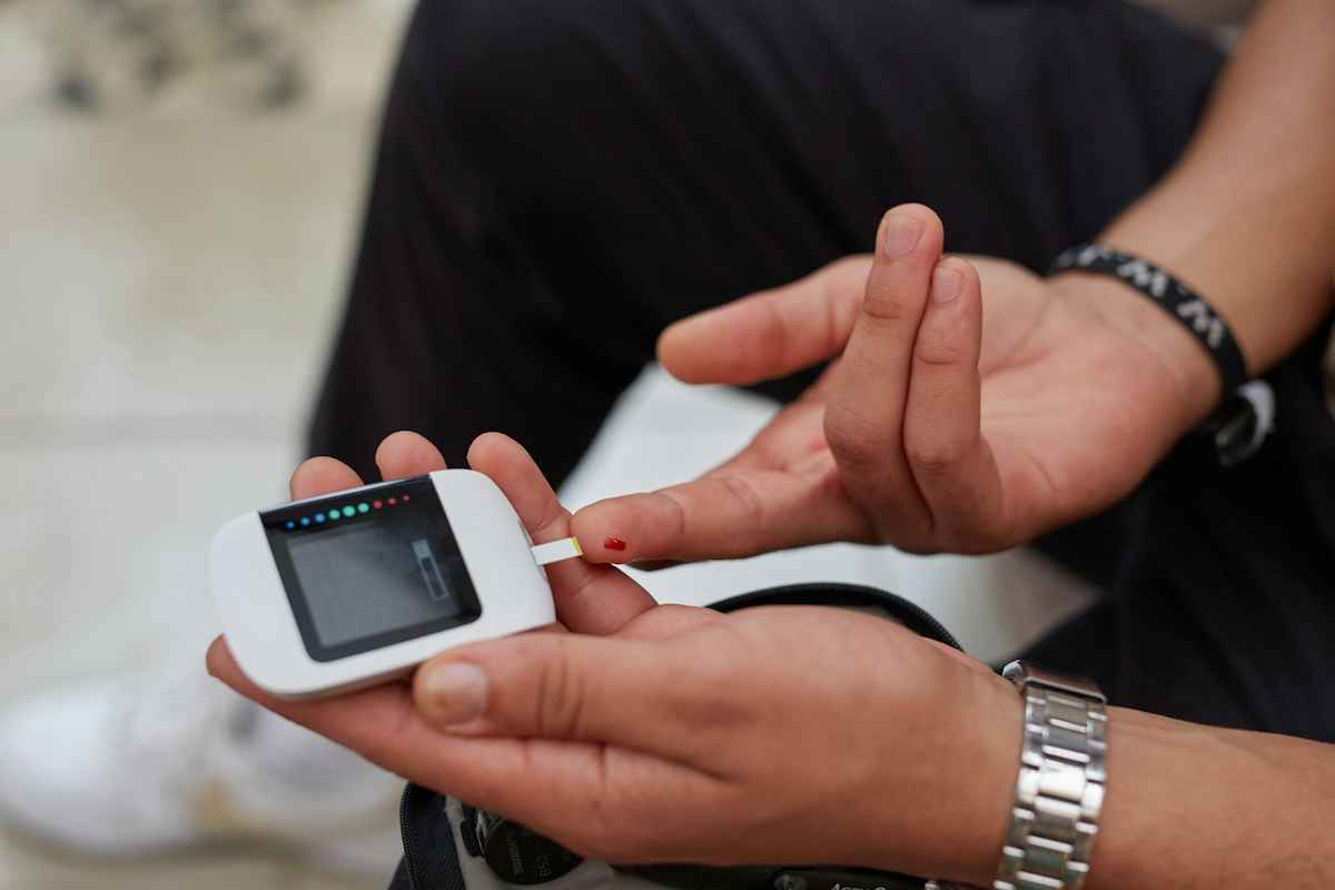
Continuous glucose monitoring (CGM) technology has achieved a transformative paradigm shift in modern health technology, expanding from a specialized, life-saving clinical medical device engineered for patients with Type 1 and advanced insulin-requiring Type 2 diabetes into a widespread consumer digital biomarker platform. Millions of metabolic health enthusiasts, endurance athletes, corporate executives, and non-diabetic individuals now wear subcutaneous biosensors to quantify real-time interstitial glucose fluctuations, evaluate dietary responses, and optimize systemic cardiometabolic health.
This rapid democratization of biosensing technology has ignited vigorous scientific debate within clinical endocrinology, preventive cardiology, and metabolic physiology. Proponents argue that continuous biofeedback enables non-diabetic individuals to identify occult postprandial glucose excursions, mitigate subclinical insulin resistance decades before diagnostic elevations in glycated hemoglobin (HbA1c) occur, and personalize nutritional intake based on unique glycemic responses. Conversely, critics highlight the lack of long-term randomized clinical trial outcomes proving hard cardiovascular risk reduction, potential sensor over-interpretation, and the psychological perils of hyper-vigilant food anxiety and orthorexic eating behaviors.
This comprehensive clinical intelligence treatise provides an exhaustive, evidence-based evaluation of continuous glucose monitoring in non-diabetic populations. We examine sensor electrochemistry and the biophysics of interstitial-to-capillary fluid delay times, analyze standardized glycemic variability metrics including Mean Amplitude of Glycemic Excursions (MAGE) and Continuous Overlapping Net Glycemic Action (CONGA), dissect the physiological modulators of postprandial glucose flux, and assess clinical trial evidence regarding long-term cardiometabolic efficacy and emerging over-the-counter biosensor regulations.
The Transposition of CGM: From Type 1 Diabetes to General Metabolic Wellness
Continuous glucose monitoring was conceived, engineered, and clinically approved as a specialized medical technology designed to address the volatile glycemic instability of Type 1 diabetes mellitus. For individuals with absolute pancreatic beta-cell destruction, real-time interstitial glucose tracking, integrated with automated trend arrows and threshold alarms, transformed diabetes care by reducing severe hypoglycemia (blood glucose < 54 mg/dL), preventing diabetic ketoacidosis, and optimizing insulin dosing.
However, over the past five years, advances in sensor miniaturization, factory calibration, extended wear durations (14 to 15 days), and direct Bluetooth smartphone integration have dramatically reduced barrier-to-entry friction. Concurrently, venture-backed digital health startups began repurposing commercial CGM hardware for healthy, non-diabetic consumer demographics under the banner of proactive metabolic optimization.
The clinical rationale underlying this expansion is rooted in the recognition of a widespread, underdiagnosed epidemic of insulin resistance and metabolic syndrome. Conventional diagnostic markers – specifically fasting plasma glucose and glycated hemoglobin (HbA1c) – are notoriously insensitive late-stage indicators of metabolic decay.
Pancreatic beta-cells can hypersecrete insulin for decades to maintain normal fasting glucose (< 100 mg/dL) and normal HbA1c (< 5.7 percent) despite severe peripheral insulin resistance. Continuous glucose monitoring exposes transient, high-amplitude postprandial glucose spikes and nocturnal dysglycemia that remain completely hidden on standard annual laboratory blood draws.
Consequently, CGM has evolved from an acute therapeutic management device into an exploratory investigative tool for early metabolic surveillance and preventative lifestyle intervention.
Sensor Electrochemistry: Glucose Oxidase, Platinum Electrodes, and Interstitial Dynamics
Understanding the physiological data generated by wearable CGM sensors requires examining the underlying analytical electrochemistry that translates biochemical glucose molecules into digital microampere electrical signals.
Commercial CGM sensors (such as Abbott FreeStyle Libre and Dexcom G6/G7) consist of a flexible, miniaturized polymer filament (typically 5 to 7 millimeters in length and less than 0.4 millimeters in diameter) that is inserted into the subcutaneous adipose tissue using a spring-loaded sterile applicator. The filament surface is coated with an immobilized enzyme layer containing glucose oxidase (GOx) encapsulated within a biocompatible, semi-permeable polyurethane or hydrogel membrane.
As interstitial glucose molecules diffuse across the outer semi-permeable membrane, glucose oxidase catalyzes the oxidation of beta-D-glucose into D-glucono-1,5-lactone and reduced flavin adenine dinucleotide (FADH2). In the presence of ambient dissolved oxygen, FADH2 is re-oxidized back to FAD, producing hydrogen peroxide (H2O2).
The generated hydrogen peroxide diffuses to an underlying platinum-iridium working electrode held at a positive polarization potential (+0.6 to +0.7 Volts relative to an Ag/AgCl reference electrode). At the platinum surface, hydrogen peroxide undergoes electrochemical oxidation: H2O2 -> O2 + 2H+ + 2e-. The liberated electrons generate an electrical current directly proportional to the concentration of glucose in the surrounding interstitial fluid.
Onboard microprocessors sample this current at intervals of 1 to 5 minutes, applying proprietary polynomial filtering algorithms to eliminate electronic noise and convert electrical nanoamperes into estimated glucose values reported in milligrams per deciliter (mg/dL) or millimoles per liter (mmol/L).
Interstitial Lag Time: Physiological Gradients Between Capillary Blood and Tissues
A fundamental physiological principle that every CGM user and clinician must understand is that wearable sensors do not measure blood glucose directly; they measure the glucose concentration of the subcutaneous interstitial fluid (ISF) bath surrounding adipocytes and dermal fibroblasts.
Glucose enters the interstitial space by passively diffusing down a concentration gradient across the fenestrations of capillary endothelial cells. During periods of stable, resting metabolic equilibrium (such as overnight fasting), interstitial glucose concentration equilibrates closely with capillary blood glucose (interstitial-to-blood ratio ~ 0.95 to 1.0).
However, during periods of rapid glycemic flux – such as following the rapid ingestion of refined carbohydrates or during intense physical exercise – a significant physiological ‘lag time’ develops between intravascular blood and subcutaneous interstitial compartments. This lag time consists of two additive components: a physiological transit delay (the time required for glucose to diffuse across endothelial walls and through extracellular matrix ground substance) and a biochemical sensor delay (the time required for glucose to permeate the sensor membrane and react enzymatically).
Extensive clinical validation trials establish that the total physiological lag time in subcutaneous adipose tissue ranges between 5 and 15 minutes. Consequently, when blood glucose is rising rapidly after a meal, the CGM sensor reading will temporarily lag below the true capillary blood fingerstick value; conversely, when glucose is falling rapidly, the sensor will read higher than true blood glucose.
Failing to account for interstitial lag time often leads to unwarranted anxiety or improper nutritional compensation in non-diabetic users who misinterpret normal physiological diffusion delays as sensor inaccuracy.
Standardized Metrics of Glycemic Control: TIR, Mean Glucose, and GMI
In clinical diabetology, continuous glucose monitoring data is standardized through the international consensus Ambulatory Glucose Profile (AGP), which synthesizes 14 consecutive days of continuous sensor readings into validated clinical metrics.
The cornerstone metric is Time in Range (TIR), defined as the percentage of time spent within the target glucose corridor of 70 to 180 mg/dL (3.9 to 10.0 mmol/L). In patients with diabetes, clinical guidelines established by the American Diabetes Association (ADA) set a therapeutic goal of TIR > 70 percent, which correlates strongly with reduced microvascular complications (retinopathy, nephropathy, and neuropathy).
In healthy, non-diabetic populations, physiological glycemic control is significantly tighter. Landmark observational studies monitoring healthy non-diabetic cohorts reveal that healthy individuals naturally spend greater than 95 to 97 percent of their time within the tighter euglycemic corridor of 70 to 140 mg/dL, with less than 1 percent of time spent below 70 mg/dL.
Other core AGP parameters include Mean Glucose (the mathematical average of all recorded readings) and the Glucose Management Indicator (GMI), an estimated HbA1c calculation derived from mean glucose: GMI (%) = 3.31 + 0.02392 * [Mean Glucose in mg/dL]. In healthy individuals, 24-hour mean interstitial glucose typically averages between 90 and 105 mg/dL, corresponding to a GMI between 5.46 and 5.82 percent.
While these consensus metrics are indispensable for managing diabetes, applying diabetic thresholds (70-180 mg/dL) to non-diabetic populations is clinically inappropriate, requiring the adoption of refined glycemic variability indices to assess subtle metabolic variations.
Quantifying Glycemic Variability: MAGE, CONGA, and J-Index Mathematical Significance
Glycemic variability (GV) quantifies the amplitude, frequency, and duration of glucose oscillations across the 24-hour circadian day. Emerging evidence in vascular biology indicates that acute, high-amplitude glycemic swings inflict greater endothelial shear stress and oxidative damage than sustained, stable moderate hyperglycemia.
The gold-standard mathematical metric for assessing within-day intraday glycemic variability is the Mean Amplitude of Glycemic Excursions (MAGE), developed by Service and colleagues. MAGE is calculated by measuring the vertical height of all glucose excursions (from nadir to peak, or peak to nadir) that exceed one standard deviation (1 SD) of the 24-hour mean glucose, averaging the amplitudes of these qualifying swings. In healthy, metabolically flexible non-diabetics, MAGE remains below 28 to 35 mg/dL; values exceeding 40 to 50 mg/dL signal early loss of first-phase insulin secretion.
Another sophisticated time-series metric is Continuous Overlapping Net Glycemic Action (CONGA), which measures the standard deviation of the difference between an observation and another observation recorded n hours earlier (e.g., CONGA-1 for 1-hour intervals, CONGA-2 for 2-hour intervals). CONGA evaluates within-day rhythmic stability without requiring arbitrary peak-and-trough definition.
The J-Index combines mean glucose level and glucose variability into a single composite mathematical score: J = 0.001 * (Mean + SD)^2. In healthy individuals, J-Index values typically range between 10 and 20, whereas values > 30 indicate metabolic dysregulation.
Additionally, the Coefficient of Variation (%CV = [SD / Mean] * 100) serves as a normalized, unit-less metric of glycemic stability. International consensus defines stable glycemic control as %CV <= 36 percent. Healthy non-diabetics consistently exhibit tight %CV values between 15 and 22 percent.
Postprandial Glucose Dynamics in Normoglycemic Adults: Redefining Normal
One of the most profound clinical revelations generated by consumer CGM studies has been the re-evaluation of what constitutes a ‘normal’ postprandial glucose excursion in healthy, non-diabetic individuals.
Historically, classical medical textbook teaching asserted that healthy humans maintain blood glucose strictly within a narrow window between 70 and 140 mg/dL, rarely exceeding 140 mg/dL even following large meals. However, landmark clinical investigations conducted at Stanford University by Hall and colleagues equipped non-diabetic individuals with CGMs and exposed them to standardized mixed meals and oral glucose tolerance tests.
The researchers discovered that over 80 percent of healthy, non-diabetic participants experienced transient postprandial glucose spikes into the prediabetic (> 140 mg/dL) and even diabetic (> 200 mg/dL) ranges following common breakfast foods (such as cornflakes with milk or bagels). The duration of these excursions was typically brief (15 to 45 minutes), with glucose rapidly clearing back below 100 mg/dL secondary to vigorous pancreatic beta-cell insulin compensation.
Based on continuous glycemic patterns, the researchers stratified non-diabetic individuals into three distinct ‘glucotypes’: low, moderate, and severe variability. Individuals in the severe variability glucotype exhibited high glycemic volatility and frequent subclinical spikes despite having completely normal fasting glucose and normal HbA1c.
This confirms that transient postprandial spikes above 140 mg/dL occur naturally in healthy individuals depending on meal composition, challenging the simplistic assumption that every post-meal spike indicates underlying pathology.
Food Sequencing and Macronutrient Co-Ingestion: Fiber, Protein, and Fat Kinetics
Continuous glucose monitoring has empirically validated decades of gastrointestinal physiological research demonstrating that the sequence in which macronutrients are ingested exerts a profound impact on postprandial glycemic excursions.
When refined carbohydrates are ingested in isolation on an empty stomach, rapid gastric emptying delivers high concentrations of monosaccharides into the duodenum. Rapid intestinal absorption via sodium-glucose cotransporter 1 (SGLT1) produces a steep, high-amplitude arterial glucose spike accompanied by an exaggerated surge in compensatory insulin secretion, frequently precipitating reactive hypoglycemia (postprandial dip below baseline).
Conversely, clinical sequencing trials demonstrate that consuming soluble viscous fiber (such as vegetables, salads, or legumes) and high-quality protein (poultry, fish, tofu, whey) 10 to 15 minutes prior to carbohydrate ingestion significantly flattens the postprandial glucose curve. Soluble fiber forms a viscous polysaccharide gel matrix within the stomach and small intestine, trapping digestive enzymes and slowing carbohydrate hydrolysis.
Simultaneously, the arrival of peptides and free amino acids in the duodenum stimulates enteroendocrine K and L cells to secrete incretin hormones: glucose-dependent insulinotropic polypeptide (GIP) and glucagon-like peptide-1 (GLP-1). GLP-1 activates vagal afferents that decelerate gastric motility (gastric emptying delay), slowing the mechanical release of chyme into the absorptive small intestine.
Furthermore, co-ingesting dietary fats delays gastric emptying via cholecystokinin (CCK) release; however, high-saturated-fat co-ingestion can prolong postprandial hyperglycemia into late hours secondary to transient, lipid-induced peripheral insulin resistance (Randle cycle fatty acid oxidation competition).
Circadian Insulin Sensitivity: Melatonin Antagonism and Late-Night Meal Spikes
The human endocrine system exhibits profound circadian rhythmicity, wherein systemic insulin sensitivity and glucose tolerance fluctuate predictably across the 24-hour solar cycle, governed by the central circadian clock in the suprachiasmatic nucleus (SCN) and peripheral clocks in pancreatic islets, liver, skeletal muscle, and adipose tissue.
Systemic insulin sensitivity is highest during the morning and early afternoon waking hours, coinciding with peak physical activity and circadian upregulation of skeletal muscle GLUT4 glucose transporter translocation. Identical meals consumed at 8:00 AM versus 8:00 PM produce radically divergent glycemic excursions: evening meals produce postprandial glucose peaks that are 20 to 50 percent higher and require double the insulin exposure to clear.
A major molecular driver of nocturnal insulin resistance is the pineal hormone melatonin. As evening darkness approaches, circulating melatonin concentrations surge to promote sleep. Melatonin binds to high-affinity melatonin receptors (MT1 and MT2) expressed on the surface of pancreatic beta-cells. Activation of MT1/MT2 receptors couples to inhibitory Gi proteins, reducing intracellular cAMP and blunting glucose-stimulated insulin secretion (GSIS).
This evolutionary mechanism serves to prevent hypoglycemia during the overnight fast; however, when individuals consume carbohydrate-rich meals late in the evening while melatonin levels are elevated, insulin secretion is blunted and delayed, causing sustained, elevated postprandial hyperglycemia that persists throughout early sleep cycles.
CGM data provides users with striking visual proof of this circadian divergence, encouraging adherence to early time-restricted feeding (eTRF) windows that align caloric intake with biological insulin sensitivity.
Exercise-Induced Glycemic Fluctuations: Aerobic Base vs High-Intensity Spikes
Physical exercise exerts profound, multi-phasic effects on systemic glucose dynamics, producing distinct CGM trajectories that frequently confuse non-diabetic users who expect exercise to universally lower blood sugar.
During low- to moderate-intensity steady-state aerobic exercise (Zone 2 conditioning, jogging, brisk walking, cycling below lactate threshold 1), skeletal muscle contractions stimulate insulin-independent glucose uptake. Mechanical tension and AMP-activated protein kinase (AMPK) activation drive the translocation of GLUT4 storage vesicles from intracellular pools to the sarcolemma, accelerating glucose clearance from the bloodstream.
Because hepatic gluconeogenesis and glycogenolysis closely match peripheral muscular glucose consumption during moderate aerobic exercise, CGM readings typically exhibit gentle, steady declines or remain rock-stable within euglycemic boundaries.
In stark contrast, high-intensity interval training (HIIT), heavy resistance training, and all-out sprint efforts performed above the anaerobic threshold trigger massive surges in systemic catecholamines (epinephrine and norepinephrine) alongside elevations in cortisol and glucagon. Epinephrine stimulates hepatic beta-2 adrenergic receptors, unleashing rapid hepatic glycogenolysis and gluconeogenesis.
Under intense anaerobic exertion, hepatic glucose production outpaces skeletal muscle glucose uptake by two- to three-fold. Consequently, non-diabetic athletes frequently observe a sharp, transient glucose spike (often reaching 150 to 180 mg/dL) during or immediately following intense interval sessions. This exercise-induced hyperglycemic spike is a completely benign, normal physiological manifestation of catecholaminergic fuel mobilization, not a sign of metabolic dysfunction.
Psychological and Physiological Stress: Adrenal Cortisol and Sympathetic Hepatic Output
Systemic glucose homeostasis is exquisitely sensitive to acute and chronic psychological stress, mediated through the neuroendocrine axes of the Sympathetic-Adreno-Medullary (SAM) system and the Hypothalamic-Pituitary-Adrenal (HPA) axis.
Under perceived psychological threat, performance anxiety, or emotional panic, the amygdala stimulates the paraventricular nucleus of the hypothalamus to release corticotropin-releasing hormone (CRH), triggering anterior pituitary adrenocorticotropic hormone (ACTH) secretion, which stimulates the adrenal cortex to release cortisol.
Simultaneously, the sympathetic nervous system triggers adrenal medullary epinephrine secretion. Cortisol and epinephrine act synergistically to mobilize energy reserves: stimulating hepatic gluconeogenesis, activating hormone-sensitive lipase in adipose tissue to release free fatty acids, and inducing acute peripheral insulin resistance in skeletal muscle by inhibiting insulin receptor substrate-1 (IRS-1) phosphorylation.
On continuous glucose monitors, non-diabetic individuals experiencing acute psychological stress (such as public speaking, intense corporate negotiations, or vehicular traffic) frequently display unprovoked glycemic elevations (glucose drifting from 85 mg/dL up to 120-140 mg/dL) in the total absence of food intake.
Chronic, unmitigated psychological stress sustains elevated basal cortisol concentrations, leading to persistent nocturnal dysglycemia, blunted morning insulin sensitivity, and gradual expansion of visceral adiposity.
Sleep Architecture and Nocturnal Glucose Homeostasis: Dawn vs Somogyi Dynamics
Continuous glucose monitoring provides an unprecedented, continuous window into nocturnal metabolic homeostasis, illuminating the profound biological dialogue between sleep architecture and endocrine regulation.
During normal, consolidated nocturnal sleep, metabolic rate decreases by 10 to 15 percent. During Slow-Wave Sleep (SWS, Stage N3), cerebral glucose utilization declines, parasympathetic vagal tone peaks, and circulating glucose concentrations remain extraordinarily stable, typically hovering in a flat, tranquil line between 75 and 95 mg/dL.
In the early pre-dawn hours (typically between 4:00 AM and 7:00 AM), the body prepares for daytime waking through the physiological ‘Dawn Phenomenon’. Pulsatile surges of growth hormone, accompanied by morning cortisol rises and sympathetic activation, stimulate hepatic gluconeogenesis and transiently decrease peripheral insulin sensitivity, causing a mild, natural rise in waking glucose (typically 5 to 15 mg/dL) before breakfast.
Furthermore, CGM sensors frequently record deceptive nocturnal ‘dips’ below 60 or 55 mg/dL that trigger alarming low-glucose alerts in non-diabetic users. In the vast majority of non-diabetic cases, these nocturnal dips are ‘compression artifacts’ caused by sleeping directly on top of the sensor. External mechanical pressure compresses the microvasculature around the sensor filament, halting local capillary blood flow and causing interstitial glucose depletion while true systemic blood glucose remains perfectly normal.
Conversely, fragmented sleep, nocturnal obstructive sleep apnoea (OSA) hypoxemic arousals, and chronic sleep deprivation disrupt nocturnal slow-wave stability, unleashing sympathetic surges that produce jagged nocturnal glycemic profiles and elevate next-morning fasting insulin levels.
Cardiovascular and Endothelial Consequences of Postprandial Spikes: ROS and Glycocalyx
A central premise motivating non-diabetic CGM use is the hypothesis that transient postprandial glucose excursions, even within subclinical thresholds, inflict cumulative microvascular and endothelial damage that accelerates atherosclerosis.
In experimental vascular biology, acute glucose spikes have been demonstrated to exert direct cytotoxic effects on human vascular endothelial cells. When endothelial cells are exposed to fluctuating glucose concentrations, the sudden intracellular influx of glucose overwhelms the mitochondrial electron transport chain.
Complex III becomes electron-saturated, generating massive quantities of superoxide radical anions (O2-). Mitochondrial superoxide inhibits glyceraldehyde-3-phosphate dehydrogenase (GAPDH), shunting glycolytic intermediates into four deleterious vascular damage pathways: the polyol (sorbitol) pathway, advanced glycation end-product (AGE) crosslinking, protein kinase C (PKC) activation, and the hexosamine pathway.
Furthermore, acute glucose spikes degrade the endothelial glycocalyx – the delicate, gel-like glycoprotein mesh lining the luminal surface of blood vessels that protects against platelet adhesion and maintains vascular nitric oxide production. Within two hours of an acute glycemic spike, circulating concentrations of shed syndecan-1 and hyaluronan elevate, indicating glycocalyx barrier disruption.
However, whether the transient, physiological postprandial spikes observed in healthy individuals with intact antioxidant systems and normal insulin clearance cause meaningful, irreversible clinical cardiovascular disease remains an active subject of ongoing long-term epidemiological investigation.
Dietary Bio-Individuality: Microbiome Composition and Personalized Nutrition
A seminal scientific milestone that catalyzed the modern consumer CGM movement was the groundbreaking 2015 personalized nutrition study conducted by Zeevi, Segal, and colleagues at the Weizmann Institute of Science, published in Cell.
The Israeli researchers continuously monitored the interstitial glucose of 800 non-diabetic participants consuming thousands of standardized and everyday meals over a full week. The study revealed astonishing bio-individuality in postprandial glycemic responses to identical foods: while some individuals exhibited steep, high-amplitude glucose spikes after consuming white bread but flat responses to white rice, other participants showed the exact opposite glycemic trajectory.
The researchers demonstrated that universal nutritional guidelines and simplistic carbohydrate-counting metrics (such as the Glycemic Index) fail to accurately predict an individual’s personal glycemic response because postprandial kinetics are governed by complex personal biological variables: body mass index, insulin sensitivity, physical activity timing, sleep debt, and most crucially, the taxonomic composition and metabolic capacity of the gut microbiome.
By training machine learning gradient-boosting algorithms on multi-omic participant data – including 16S rRNA gut microbiome profiles, continuous glucose metrics, and dietary logs – the researchers successfully predicted personalized postprandial responses and engineered personalized dietary interventions that significantly blunted postprandial glycemic spikes.
This demonstrated that continuous biofeedback can help individuals identify specific dietary triggers that provoke exaggerated glycemic reactions, moving nutrition science away from generic population dogmas toward personalized metabolic management.
Behavioral Psychology and Eating Disorders: Orthorexia, Anxiety, and Glucose Obsession
While the physiological data provided by continuous glucose monitors can empower proactive health behaviors, clinical psychologists and eating disorder specialists have documented significant unintended behavioral and psychiatric harms arising from non-diabetic CGM utilization.
In healthy individuals without diabetes, continuous access to real-time, minute-by-minute biological data can precipitate profound health anxiety, obsessive-compulsive checking behaviors, and the emergence of orthorexia nervosa – a pathological obsession with righteous, clean eating.
Because consumer health apps frequently gamify glucose tracking – rewarding users with ‘green scores’ for flat lines and punishing any postprandial elevation with alarming red spikes – non-diabetic users often develop irrational food phobias, viewing normal physiological glucose excursions as catastrophic metabolic failures. Users frequently begin eliminating nutrient-dense, health-promoting whole foods (such as fresh fruits, root vegetables, legumes, and whole grains) simply because they induce normal, transient postprandial elevations.
Furthermore, hyper-vigilance over glucose metrics can decouple individuals from natural intuitive hunger and satiety signals, replacing internal interoceptive awareness with external digital validation.
Clinical endocrinologists emphasize that CGM data should be interpreted with psychological balance: a completely flat glucose line is neither biologically necessary nor indicative of optimal health, and the emotional stress generated by obsessive monitoring can produce cortisol-mediated glycemic elevations that exceed the impact of the food itself.
Device Accuracy and Interfering Substances: MARD Benchmarks and Chemical Bias
Accurately interpreting CGM data requires an understanding of analytical sensor accuracy and the chemical substances that can induce spurious false readings.
The standard clinical metric for assessing CGM accuracy is the Mean Absolute Relative Difference (MARD), calculated as the average percentage difference between continuous sensor readings and simultaneous reference laboratory blood glucose measurements (such as YSI 2300 STAT glucose analyzers): MARD = (1/N) * sum(|CGM_i – Ref_i| / Ref_i) * 100. Lower MARD percentages indicate superior analytical accuracy; modern commercial sensors achieve MARD values between 8.2 and 9.5 percent in diabetic cohorts.
However, in healthy non-diabetic populations whose glucose fluctuates within a tighter euglycemic range (70 to 120 mg/dL), small absolute variations (such as a sensor reading 75 mg/dL when true blood is 85 mg/dL) generate disproportionately high relative percentage errors, artificially inflating MARD.
Furthermore, several common chemical substances can electrochemically interfere with sensor electrodes. Acetaminophen (paracetamol) is electrochemically active at the polarization potential of first-generation platinum electrodes, undergoing oxidation and producing false, dramatic spikes in estimated glucose. While newer sensors utilize protective screening membranes that block acetaminophen, high-dose intravenous or oral Vitamin C (ascorbic acid, > 1,000 mg) can cause false elevations on certain sensor models.
Similarly, severe dehydration, peripheral hypoperfusion, and cutaneous hypothermia reduce subcutaneous capillary blood flow, leading to falsely depressed interstitial glucose readings.
Clinical Trial Evidence: Long-Term Cardiometabolic Outcomes in Non-Diabetics
Despite enthusiastic commercial marketing and widespread adoption among biohackers and wellness enthusiasts, a rigorous examination of the peer-reviewed medical literature reveals an absence of large-scale, prospective randomized controlled trials demonstrating that CGM use in healthy, non-diabetic adults improves hard clinical endpoints.
While short-term crossover studies confirm that wearing a CGM can induce positive behavioral modifications – such as increasing post-meal walking, reducing consumption of refined sugary beverages, and improving meal sequencing – there is currently no prospective clinical trial data proving that flattening postprandial glucose curves in normoglycemic individuals reduces the long-term incidence of cardiovascular disease, myocardial infarction, stroke, dementia, or all-cause mortality.
Major clinical professional societies, including the American Diabetes Association (ADA) and the Endocrine Society, have issued clinical position statements emphasizing that continuous glucose monitoring is not medically indicated or clinically recommended for individuals without diagnosed diabetes or documented hypoglycemia disorders outside of investigational research protocols.
Endocrinologists caution that for individuals with normal fasting glucose, normal HbA1c, and absence of metabolic syndrome criteria, investing thousands of dollars in commercial CGM subscriptions provides questionable clinical value compared to established lifestyle fundamentals: regular resistance exercise, balanced Mediterranean dietary patterns, quality sleep, and routine lipid screening.
Regulatory Landscapes and Over-the-Counter Biosensor Evolution
The regulatory landscape governing continuous glucose monitoring is undergoing a historic evolution, transitioning from strict prescription-only medical device status toward cleared over-the-counter (OTC) consumer wellness biosensors.
In early 2024, the United States Food and Drug Administration granted historic 510(k) clearance for the first over-the-counter continuous glucose monitoring systems – including Dexcom’s Stelo and Abbott’s Lingo and Libre Rio systems – specifically indicated for non-diabetic adults and individuals with Type 2 diabetes who do not manage their condition with insulin.
These next-generation consumer biosensors feature modified software interfaces tailored specifically for wellness applications: eliminating high-acuity hypoglycemia alarms, extending sensor wear duration to 15 days, and presenting data through intuitive metabolic scores rather than complex clinical ambulatory glucose profile graphs.
This regulatory milestone establishes a new category of consumer metabolic wearables, positioning continuous glucose tracking alongside smartwatches and fitness rings. However, regulatory agencies emphasize that consumer OTC biosensors are cleared for general wellness and behavioral lifestyle tracking, and cannot be utilized for medical diagnosis or therapeutic drug titration without licensed medical supervision.
To provide endocrinologists, primary care physicians, sports scientists, and digital health practitioners with an evidence-based clinical matrix, the following comparative framework outlines the analytical definitions, physiological drivers, target reference ranges, and clinical interpretations of standard glycemic metrics across both healthy non-diabetic and diabetic populations. Each metric is classified according to its diagnostic utility, mathematical derivation, and behavioral significance.
Applying this structured matrix ensures that clinicians and health consumers interpret wearable biosensor data with scientific rigor, preventing misinterpretation of normal physiological excursions while identifying genuine metabolic warning signs.
| Glycemic Metric / Parameter | Mathematical Derivation / Units | Healthy Non-Diabetic Target | Diabetic Consensus Target | Primary Clinical Interpretation |
|---|---|---|---|---|
| Time in Range (TIR) | Percentage of readings within target corridor (%) | > 95% in 70 – 140 mg/dL corridor | > 70% in 70 – 180 mg/dL corridor | Gold standard for overall glycemic control; strongly correlates with microvascular risk |
| Mean 24h Interstitial Glucose | Mathematical average of all 24h readings (mg/dL) | 85 – 105 mg/dL (4.7 – 5.8 mmol/L) | < 154 mg/dL (corresponding to HbA1c < 7.0%) | Reflects chronic basal glucose exposure and hepatic glucose output |
| Mean Amplitude Excursions (MAGE) | Average height of swings exceeding 1 SD of mean (mg/dL) | < 35 mg/dL | Variable; clinical goal < 60 mg/dL | Pure measure of postprandial volatility; reflects first-phase insulin response |
| Coefficient of Variation (%CV) | (Standard Deviation / Mean Glucose) * 100 (%) | 15 – 22% | <= 36% (target for stable control) | Normalized index of glycemic stability; values > 36% indicate severe volatility |
| Time Below Range (TBR) | Percentage of readings < 70 mg/dL (Level 1 hypo) | < 1 - 2% (often compression artifacts) | < 4% (< 70 mg/dL); < 1% (< 54 mg/dL) | Critical safety marker; in non-diabetics, nocturnal drops are usually compression artifacts |
The operational guidelines delineated in the table above emphasize that interpreting continuous glucose metrics requires population-specific clinical context. While a Time in Range of 75 percent represents successful clinical management for an individual with insulin-dependent diabetes, it would signal significant metabolic dysregulation in a healthy non-diabetic adult.
Furthermore, understanding that healthy individuals naturally exhibit transient excursions above 140 mg/dL following carbohydrate-dense meals prevents unnecessary dietary restriction and eliminates false self-diagnoses of prediabetes.
Frequently Asked Questions About CGM in Non-Diabetics
Do healthy non-diabetic people experience glucose spikes above 140 mg/dL?
Yes. Landmark studies using CGMs on healthy non-diabetic adults show that over 80 percent of individuals experience transient glucose spikes above 140 mg/dL, and occasionally above 180-200 mg/dL, after consuming high-carbohydrate meals like cereal or bagels. These brief spikes are normal physiological responses that typically clear rapidly within 30 to 45 minutes.
What is the difference between interstitial glucose and blood glucose?
Blood glucose is measured directly in capillary or venous blood, whereas wearable CGMs measure glucose in the subcutaneous interstitial fluid surrounding fat cells. Because glucose must diffuse from capillaries into tissues, there is a normal physiological lag time of 5 to 15 minutes between blood and interstitial readings during rapid glucose changes.
Why does my glucose sensor read low when I am sleeping?
Most nocturnal low-glucose alerts in non-diabetics are ‘compression artifacts’. Sleeping directly on the sensor mechanically compresses local skin capillaries, temporarily stopping local blood flow and causing interstitial glucose levels around the sensor to drop artificially, while true blood glucose remains perfectly normal.
Can wearing a CGM help someone lose weight?
CGMs can assist weight management indirectly by increasing awareness of food choices, discouraging sugary refined snacks, and encouraging post-meal physical activity. However, a CGM does not burn calories or guarantee weight loss; calorie balance and diet quality remain the fundamental drivers of fat loss.
Why does high-intensity exercise cause blood sugar to spike?
Intense anaerobic exercise (such as sprinting or heavy lifting) triggers a surge of stress hormones, particularly adrenaline and cortisol. These hormones stimulate the liver to release stored glycogen into glucose faster than working muscles can absorb it, causing a temporary, harmless blood sugar spike that normalizes quickly after the workout.
How does food sequencing affect post-meal glucose?
Consuming vegetables (fiber) and protein before carbohydrates significantly flattens post-meal glucose spikes. Soluble fiber slows gastric emptying and carbohydrate absorption, while protein stimulates incretin hormones (GLP-1) that prime insulin release, resulting in smoother glycemic curves.
Can wearing a CGM cause eating disorders or food anxiety?
Yes. Clinical psychologists have noted that continuous biofeedback can provoke health anxiety, obsessive calorie-counting, and orthorexia nervosa (an obsession with clean eating). Users may begin fearing healthy foods like fruit or sweet potatoes simply because they cause a normal, temporary glucose rise.
What is Mean Amplitude of Glycemic Excursions (MAGE)?
MAGE is a mathematical metric that measures the average height of significant glucose swings (exceeding one standard deviation of the daily mean). It reflects postprandial glucose volatility and beta-cell response speed, with normal non-diabetic values remaining below 35 mg/dL.
What are the new over-the-counter (OTC) CGMs approved by the FDA?
In 2024, the FDA cleared the first over-the-counter CGMs, such as Dexcom Stelo and Abbott Lingo, for adults without diabetes. These wellness biosensors track daily metabolic trends and do not require a doctor’s prescription, but they are not intended for diabetes diagnosis or insulin dosing.
Is there evidence that CGM improves long-term health in healthy people?
Currently, there are no long-term randomized clinical trials proving that wearing a CGM prevents heart attacks, strokes, dementia, or premature death in healthy non-diabetic individuals. Major medical organizations like the American Diabetes Association do not recommend routine CGM use for healthy adults outside of clinical studies.
Clinical Perspectives and Future Directions in Consumer Biosensing
Continuous glucose monitoring represents a technological marvel that has permanently altered the landscape of preventative health, metabolic science, and personalized medicine. By rendering invisible metabolic physiology instantly visible, CGM empowers individuals to observe the direct consequences of culinary choices, physical exercise, nocturnal sleep quality, and psychological stress on systemic fuel homeostasis.
However, the clinical translation of this technology into non-diabetic wellness requires scientific nuance and psychological vigilance. Biosensor data must be contextualized within established physiological boundaries, recognizing that natural glycemic oscillations are an inherent feature of human biology rather than a pathological failure. When utilized thoughtfully as a temporary educational tool alongside evidence-based lifestyle foundations, continuous biosensing can serve as a powerful catalyst for enduring metabolic vitality.
For accredited institutional consensus and clinical guidance on continuous glucose monitoring and glycemic metrics, healthcare professionals and consumers are encouraged to review clinical practice statements published by the American Diabetes Association (ADA), the Endocrine Society, and the American Association of Clinical Endocrinology (AACE). Ongoing digital biomarker research is continuously indexed on PubMed National Library of Medicine, alongside global metabolic health frameworks from the World Health Organization.
